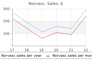
Purchase norvasc 5 mg overnight delivery
For instance arrhythmia medications buy norvasc 10 mg line, it may add carbohydrate chains to the proteins blood pressure up during pregnancy discount norvasc 2.5 mg, producing the glycoproteins talked about in section 2. Some of the Golgi vesicles become lysosomes, while others turn into secretory vesicles that migrate to the plasma membrane and fuse with it, releasing the cell product by exocytosis. This is how a cell of a salivary gland, for example, secretes mucus and digestive enzymes, and how a cell of the pituitary gland releases thyroid-stimulating hormone. The genes for hemoglobin and digestive enzymes, for example, are current but inactive in liver cells and skin cells. In the ensuing days, the hormone prolactin stimulates cells of her mammary glands to synthesize the varied components of breast milk, together with the protein casein- something her physique has by no means synthesized earlier than. The receptor triggers the activation of a regulatory protein (transcription activator) in the cytoplasm. The secretory vesicles release the casein by exocytosis, and it turns into part of the milk. Moving the chromatin over to the nuclear lamina is another method of silencing some of its genes. At step 4, there are multiple ways in which regulatory proteins can activate gene transcription. They are produced by enzymatic reactions, and enzymes are proteins encoded by genes. But to make it, a cell of the testis takes in ldl cholesterol and enzymatically converts it to testosterone. Yet a further implication of this is that genes may tremendously affect such advanced outcomes as habits, since testosterone strongly influences such behaviors as aggression and intercourse drive. Summarize the processing of a protein from the time a ribosome finishes its work to the time a protein is secreted from the cell. The hormone prolactin triggers intracellular reactions that activate a regulatory protein and lead ultimately to the secretion of casein. From pituitary 3 When testosterone is required, luteinizing hormone stimulates manufacturing of a second messenger within the cell. O 6 Testosterone is secreted from the cell and exerts varied anatomical, physiological, and behavioral e ects. More importantly, it allows a cell to reproduce one strand based on info in the other. Even at this fee, nevertheless, it might take weeks for one polymerase molecule to replicate even one chromosome. As a result, only one mistake stays for every billion base pairs replicated-a very excessive degree of replication accuracy, if not completely flawless. One purpose is that a new base sequence generally codes for a similar factor because the old one. For instance, the beta chain of hemoglobin is 146 amino acids long in each humans and horses, however 25 of those amino acids differ between the 2 species. Other mutations, however, could kill a cell, flip it cancerous, or trigger genetic defects in future generations. Clearly some amino acid substitutions are extra crucial than others, and this affects the severity of a mutation. Phases G1, S, and G2 are collectively referred to as interphase-the time between M phases. Stomach and skin cells divide quickly, whereas bone and cartilage cells divide slowly. The steadiness between cells that are actively cycling and those standing by in G0 is an important consider determining the number of cells in the body. An inability to stop cycling and enter G0 is attribute of most cancers cells (see Deeper Insight 4. During this time, a cell synthesizes proteins, grows, and carries out its preordained duties for the body. Almost all of the dialogue in this guide relates to what cells do in the G1 phase. In cultured cells called fibroblasts, which divide every 18 to 24 hours, G1 lasts 8 to 10 hours. This is the point at which the cell carries out the semiconservative replication described earlier. In G2, a cell reveals additional growth, makes extra organelles, finishes replicating its centrioles, and synthesizes enzymes that management cell division. Meiosis, nevertheless, is restricted to one purpose, the manufacturing of eggs and sperm, and is due to this fact handled in section 27. Mitosis serves all the other capabilities of cell division: � improvement of an individual, composed of some 50 trillion � � � cells, from a one-celled fertilized egg; development of all tissues and organs after start; replacement of cells that die; and repair of broken tissues. Four phases of mitosis are recognizable: prophase, metaphase, anaphase, and telophase (fig. The nuclear envelope disintegrates during prophase and releases the chromosomes into the cytosol. The centrioles start to sprout elongated microtubules called spindle fibers, which push the centrioles aside as they grow. Some spindle fibers grow towards the chromosomes and turn out to be attached to the kinetochore on all sides of the centromere (see fig. The spindle fibers then tug the chromosomes back and forth until they line up alongside the midline of the cell. The pictures show mitosis in whitefish eggs, the place chromosomes are comparatively straightforward to observe. The drawings present a hypothetical cell with only two chromosome pairs; in people, there are 23 pairs. Long microtubules reach out from every centriole to the chromosomes, and shorter microtubules type a starlike aster,eight which anchors the assembly to the inside of the plasma membrane at each finish of the cell. Each chromatid is now thought to be a separate, single-stranded daughter chromosome. One daughter chromosome migrates to every pole of the cell, with its centromere leading the way and the arms trailing behind. Migration is achieved via motor proteins within the kinetochore crawling alongside the spindle fiber as the fiber itself is "chewed up" and disassembled on the chromosomal end. Since sister chromatids are genetically equivalent, and since every daughter cell receives one chromatid from every chromosome, the daughter cells of mitosis are genetically similar. It is achieved by the motor protein myosin pulling on microfilaments of actin in the terminal web of the cytoskeleton. This creates a crease known as the cleavage furrow around the equator of the cell, and the cell eventually pinches in two. Be aware, nevertheless, that mitosis (nuclear division) can occur without cytokinesis (cellular division). This is why some cells purchase two or extra nuclei or a quantity of equivalent units of chromosomes. The activation and inhibition of cell division are subjects of intense analysis for apparent causes similar to administration of most cancers and tissue repair.
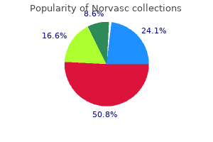
Trusted norvasc 5 mg
In capillaries and veins arteria y arteriola buy norvasc 2.5 mg with amex, the blood flows at a gentle speed with little if any pulsation as a outcome of the strain surges have been damped out by the space traveled and the elasticity of the arteries blood pressure goals 2015 norvasc 5 mg. As we grow old, our arteries become much less distensible and take in much less systolic pressure. This growing stiffness of the arteries is called arteriosclerosis6 ("hardening of the arteries"). Another contributing factor is atherosclerosis, the expansion of lipid deposits within the arterial partitions (see Deeper Insight 19. These deposits can turn out to be calcified sophisticated plaques, giving the arteries a hard, crunchy or bonelike consistency. Common blood pressures at the age of 20 are about 123/76 for males and 116/72 for females. For healthy individuals at age 70, typical blood pressures are round 145/82 and 159/85 for the two sexes, respectively. It may be a consequence of blood loss, dehydration, anemia, or different factors and is regular in individuals approaching dying. Blood strain is physiologically determined by three principal variables: cardiac output, blood volume, and resistance to move. Moving blood would exert no pressure against a vessel wall except it encountered no less than some downstream resistance. Resistance, in turn, hinges on three variables that we contemplate now: blood viscosity, vessel length, and vessel radius. Increasing distance from left ventricle Blood Viscosity the viscosity of blood stems primarily from its plasma proteins (albumin) and erythrocytes (see section 18. A deficiency of erythrocytes (anemia) or albumin (hypoproteinemia) reduces viscosity and speeds up blood flow. On the opposite hand, viscosity increases and move declines in such conditions as polycythemia and dehydration. Because of arterial elasticity and the effect of friction towards the vessel wall, all measures of blood strain decline with distance-systolic pressure, diastolic stress, pulse stress, and mean arterial pressure. In a reclining person, a powerful pulse in the dorsal artery of the foot is an effective sign of adequate cardiac output. In a healthy individual, the only vital ways of controlling peripheral resistance from second to moment are vasoconstriction, the narrowing of a vessel, and vasodilation, the widening of a vessel. Vasodilation, nevertheless, is brought about not by any muscular effort to widen a vessel, but somewhat by muscular passivity-relaxation of the smooth muscle, permitting blood stress to increase the vessel. Vasomotion is managed partly by a nucleus within the medulla oblongata of the brain called the vasomotor heart. The impact of vessel radius on blood flow stems from the friction of the transferring blood towards the vessel partitions. That is, it flows in layers- quicker near the middle of a vessel, where it encounters much less friction, and slower close to the partitions, the place it drags towards the vessel. The present could additionally be very swift in the midst of a river however fairly sluggish close to shore, the place the water encounters more friction in opposition to the riverbank and bottom. When a blood vessel dilates, a greater portion of the blood is in the midst of the stream and the common flow may be fairly swift. When the vessel constricts, extra of the blood is close to the wall and the average circulate is slower (fig. Indeed, flow (F) is proportional not merely to vessel radius (r) but to the fourth energy of radius-that is, F r4. For the sake of simplicity, contemplate a hypothetical blood vessel with a 1 mm radius when maximally constricted and a 3 mm radius when completely dilated. By the method F r4, think about how the move would change as radius changed: if r = 1 mm, then r4 = 14 = 1, and F = 1 mL/min. Blood flows extra slowly close to the vessel wall, as indicated by shorter arrows, than it does near the middle of the vessel. Each arrow may be construed as the space that a hypothetical blood cell would journey in a given period of time, varying with its distance from the vessel wall. From aorta to capillaries, velocity diminishes for 3 causes: (1) the blood has traveled a greater distance, so friction has slowed it down. The aorta has a cross-sectional area of 3 to 5 cm2, whereas the whole cross-sectional area of all the capillaries is about 4,500 to 6,000 cm2. Thus, a given volume of aortic blood is distributed over a higher complete area in the capillaries, which collectively type a wider path within the bloodstream. Just as water slows down when a slim mountain stream flows right into a lake, blood slows down as it enters pathways with a larger total area or volume. Note, nonetheless, that blood within the veins never regains the velocity it had in the massive arteries. Arterioles alone account for about half of the whole peripheral resistance of the circulatory system. However, larger arteries and veins additionally influence peripheral resistance via their own constriction and dilation. There are three ways of controlling vasomotor exercise: local, neural, and hormonal mechanisms. A single drop of epinephrine applied here triggered the arteriole to constrict to about one-third of its dilated diameter. Local Control Autoregulation is the power of tissues to regulate their own blood provide. As the bloodstream delivers oxygen and carries away the metabolites, the vessels reconstrict. In addition, platelets, endothelial cells, and the perivascular tissues secrete a variety of vasoactive chemical compounds that stimulate vasodilation under such situations as trauma, irritation, and train. The drag of blood flowing towards the endothelial cells creates a shear stress (like rubbing your palms together) that stimulates them to secrete prostacyclin and nitric oxide, which are vasodilators. Reactive hyperemia is seen when the pores and skin flushes (reddens) after a person comes in from the cold. It additionally happens in the forearm if a blood strain cuff is inflated for too long after which loosened. Over an extended time, a hypoxic tissue can enhance its personal perfusion by angiogenesis8-the progress of latest blood vessels. One cause for this is that the veins are larger than the capillaries, so they create less resistance. There is great scientific significance in determining how development factors and inhibitors management angiogenesis. Malignant tumors secrete growth elements that stimulate a dense network of vessels to develop into them and supply nourishment to the most cancers cells. Elevated blood strain Reduced blood stress Vasodilation Arteries stretched Reduced heart fee Reduced vasomotor tone Baroreceptors improve firing rate Increased vagal tone Neural Control In addition to local control, the blood vessels are beneath distant management by the central and autonomic nervous methods. The vasomotor center of the medulla oblongata exerts sympathetic control over blood vessels throughout the physique. The position of sympathetic and vasomotor tone in controlling vessel diameter is explained in section 15.
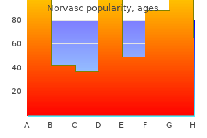
Buy norvasc 5 mg
They also can differentiate into endothelial and clean muscle cells and thus contribute to vessel progress and repair blood pressure zero gravity generic norvasc 10 mg online. Fenestrated capillaries have endothelial cells riddled with patches of filtration pores (fenestrations5) (fig prehypertension epidemiology consequences and treatment buy cheap norvasc 5 mg on-line. They allow for the fast passage of small molecules, however still retain most proteins and larger particles in the bloodstream. Fenestrated capillaries are essential in organs that interact in rapid absorption or filtration-the kidneys, endocrine glands, small gut, and choroid plexuses of the mind, for example. Sinusoids are irregular blood-filled spaces within the liver, bone marrow, spleen, and some other organs (fig. They are twisted, tortuous passageways, sometimes 30 to forty �m broad, that conform to the form of the encompassing tissue. The structures shown right here in the right carotid arteries are repeated within the left carotids. There are only two places within the circulation where this occurs- the capillaries and a few venules. We can consider these as the "enterprise end" of the cardiovascular system, as a result of all the the rest of the system exists to serve the trade processes that occur here. Capillaries are sometimes referred to as the exchange vessels of the cardiovascular system; the arterioles, capillaries, and venules are additionally referred to as the microvasculature (microcirculation). They average about 5 �m in diameter at the proximal finish (where they receive arterial blood), widen to about 9 �m on the distal finish (where they empty into a small vein), and infrequently branch alongside the way. McNutt Nonfenestrated area Nonfenestrated space Filtration pores (fenestrations) Filtration pores (fenestrations) (c) (c) four hundred nm four hundred nm Macrophage Endothelial cells Erythrocytes in sinusoid with no basal lamina, and the cells additionally frequently have especially large fenestrations by way of them. Even proteins and blood cells can pass via these pores; that is how albumin, clotting components, and other proteins synthesized by the liver enter the blood, and how newly formed blood cells enter the circulation from the bone marrow and lymphatic organs. Liver cell (hepatocyte) Microvilli Sinusoid Capillary Beds Capillaries are organized into webs referred to as capillary beds- usually 10 to 100 capillaries provided by a single arteriole or metarteriole (fig. Large gaps between the endothelial cells permit blood plasma to instantly contact the liver cells however retain blood cells in the lumen of the sinusoid. In the skeletal muscular tissues, for instance, about 90% of the capillaries have little or no blood circulate during periods of rest. During train, they obtain an ample flow whereas capillaries elsewhere-for instance, within the skin and intestines- shut down to compensate. Capillary move (perfusion) is often regulated by the dilation or constriction of arterioles upstream from the capillary beds. If most of the sphincters constrict, blood bypasses the capillaries, leaving them less perfused and even bloodless, and the blood takes a shortcut by way of the metarteriole directly to a nearby venule (fig. At relaxation, about 64% of the blood is discovered within the systemic veins as in contrast with only 13% within the systemic arteries (fig. In massive arteries, blood pressure averages 90 to one hundred mm Hg and surges to a hundred and twenty mm Hg during systole, whereas in veins it averages about 10 mm Hg. Furthermore, the blood flow within the veins is regular, somewhat than pulsating with the heartbeat just like the circulate in the arteries. They collapse when empty and thus have comparatively flattened, irregular shapes in histological sections (see fig. Capillaries 5% distal end, capillaries transition to venules, steadily adding a thin tunica media. They may drain into the distal end of a metarteriole, which then leads to a venule. What anatomical reality permits the veins to comprise a lot extra blood than the arteries do In the venous system, conversely, we find small veins merging to form larger and bigger ones as they approach the heart. We discuss with the smaller veins as tributaries, by analogy to the streams that converge and act as tributaries to rivers. Postcapillary venules are the smallest of the veins, beginning with diameters of about 10 to 20 �m. They receive blood from capillaries instantly or by the use of the distal ends of the metarterioles. Postcapillary venules are even more porous than capillaries; therefore, venules additionally change fluid with the encompassing tissues. They have a tunica media of one or two layers of smooth muscle, and a skinny tunica externa. Most veins with individual names are in this category, such because the radial and ulnar veins of the forearm and the small and nice saphenous veins of the leg. Medium veins have a tunica interna with an endothelium, basement membrane, unfastened connective tissue, and sometimes a thin inside elastic lamina. The muscle is interrupted by regions of collagenous, reticular, and elastic tissue. Many medium veins, especially within the limbs, exhibit infoldings of the tunica interna that meet in the course of the lumen, forming venous valves directed towards the heart (see fig. The upward circulate of blood in these vessels depends partly on the massaging action of skeletal muscular tissues and the power of those valves to maintain the blood from dropping down once more when the muscles relax. When the muscles surrounding a vein contract, they pressure blood via these valves. The propulsion of venous blood by muscular massaging, aided by the venous valves, is a mechanism of blood move known as the skeletal muscle pump. Varicose veins lead to part from the failure of the valves (see Deeper Insight 20. Venous sinuses are veins with especially thin walls, large lumens, and no smooth muscle. Examples embody the coronary sinus of the guts and the dural sinuses of the mind. They have a relatively skinny tunica media with solely a moderate quantity of easy muscle; the tunica externa is the thickest layer and accommodates longitudinal bundles of smooth muscle. Large veins include the venae cavae, pulmonary veins, internal jugular veins, and renal veins. Stretching pulls the cusps of the venous valves farther apart until the valves become incapable of sealing the vessel and preventing the backflow of blood. As the veins turn out to be additional distended, their walls weaken and they develop into varicose veins with irregular dilations and twisted pathways (fig. Obesity and pregnancy also promote growth of varicose veins by placing strain on large veins of the pelvic region and obstructing drainage from the decrease limbs. With much less drainage of blood, tissues of the leg and foot may become edematous and painful. Blood usually passes by way of only one community of capillaries from the time it leaves the center till the time it returns (fig. Portal techniques occur in the kidneys, join the hypothalamus to the anterior pituitary gland, and join the intestines to the liver (see section 20.
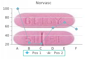
Purchase norvasc 10 mg fast delivery
However essential hypertension norvasc 2.5 mg buy discount on line, the technology of action potentials is a comparatively time-consuming course of that slows down the nerve signal on the nodes arrhythmia ablation 2.5 mg norvasc cheap free shipping. Since action potentials happen only on the nodes, this mode of conduction creates a misunderstanding that the nerve signal jumps from node to node (fig. The doors open (like the Na+ gates at a node), 20 more people get on (like Na+ flowing into the axon), and everyone has to push to the rear of the automobile to make room for them. No one passenger moves from the door to the rear, but the crowding and transfer of power from individual to particular person forces even those at the rear to transfer a little, like the sodium ions on the next node. In abstract, saltatory conduction is based on a process that is very fast in the internodal segments (transfer of energy from ion to ion), but decremental. Since most of the axon is roofed with myelin, conduction happens mainly by the quick internodal course of. This is why myelinated fibers conduct alerts much quicker (up to a hundred and twenty m/s) than unmyelinated ones (up to 2 m/s). Explain why this mechanism would block the conduction of ache signals from your enamel to your mind. Explain why myelinated fibers conduct signals a lot faster than unmyelinated fibers. All good issues should come to an end; a nerve sign soon reaches the top of an axon and can go no farther. But typically, it triggers the release of a neurotransmitter that stimulates a model new wave of electrical activity within the next cell across the synapse. The most completely studied synapse is the neuromuscular junction described in chapter eleven, but right here we contemplate synapses between two neurons. Signals arrive at the synapse by method of the presynaptic neuron, which releases a neurotransmitter. They are essential in synchronizing the activity of native suites of neurons in certain regions of the mind. The ability to do that may be a property of chemical synapses, in which neurons communicate by neurotransmitters. Chemical synapses are also the location of studying and reminiscence, the target of many prescribed drugs, and the site of motion of drugs of habit, amongst different things. A reserve pool of synaptic vesicles is located somewhat farther away from the membrane, tethered to the cytoskeleton. Its membrane does, however, have neurotransmitter receptors and ligand-gated ion channels. The presynaptic neuron may synapse with a dendrite, the soma, or the axon of a postsynaptic neuron, forming an axodendritic, axosomatic, or axoaxonic synapse, respectively (fig. For example, a spinal motor neuron is roofed with about 10,000 axon terminals from other neurons-8,000 ending on its dendrites and another 2,000 on the soma. In part of the mind known as the cerebellum, one neuron can have as many as a hundred,000 synapses. Cajal was rudely criticized for such a "preposterous" concept, however he was ultimately proved appropriate. In 1921, German pharmacologist Otto Loewi conclusively demonstrated that neurons communicate by releasing chemical compounds. He stimulated the vagus nerve of 1 frog, and its heart rate dropped as expected. He then eliminated saline from that coronary heart and squirted it onto the heart of the second frog. Loewi had found what we now call acetylcholine-the first recognized neurotransmitter. Most of them are small organic molecules which may be launched by exocytosis and bind to specific receptors on the receiving cell, but there are exceptions. Parts of the mind referred to in this table will become familiar as you examine chapter 14, and you could want to refer back to this table then to enhance your understanding of brain operate. They are synthesized as needed quite than saved in synaptic vesicles; they merely diffuse out of the axon terminal rather than being released by exocytosis; and they diffuse into the postsynaptic neuron somewhat than bind to a floor receptor. Neuropeptides are stored in secretory granules (dense-core vesicles) which would possibly be about 100 nm in diameter, twice as large as typical synaptic vesicles. Some neuropeptides additionally perform as hormones or as neuromodulators (see section 12. Some of those cause cravings for particular vitamins such as fat, protein, or carbohydrates (see part 26. Most human and different mammalian neurons can secrete two or extra neurotransmitters and can switch from one to one other beneath different circumstances. The neuropeptides are chains of amino acids, every recognized by its three-letter code. Like other local potentials, if this is robust and persistent sufficient (that is, if sufficient present makes it to the axon hillock), it opens voltage-gated ion channels within the set off zone and causes the postsynaptic neuron to hearth. When it opens, Cl� enters the cell and makes the inside much more negative than the resting membrane potential. Acetylcholine directly opens ion channels within the plasma membrane of the postsynaptic neuron. One impact is to produce an internal chemical that binds to a ligand-gated ion channel from the inside, opening the channel and depolarizing the cell. Another is to activate preexisting cytoplasmic enzymes, which may result in numerous metabolic adjustments (for instance, inducing a liver cell to break down glycogen and launch glucose into the blood). The arrival of a nerve sign at the axon terminal opens voltage-gated calcium channels. Although illustrated individually, Na+ and K+ pass in reverse directions through the identical gates. This immediately suggests a way of stopping synaptic transmission- stop adding new neurotransmitter and do away with that which is already there. The first step is achieved simply by the cessation of indicators within the presynaptic nerve fiber. The second could be achieved in the following ways, pictured as steps 6 to 8 in determine 12. A neurotransmitter or its breakdown products are reabsorbed by transport proteins within the axon terminal, removing them from the synapse and ending their stimulatory impact. Neurotransmitters or their breakdown merchandise merely diffuse away from the synapse into the nearby extracellular fluid. This is the time from the arrival of a sign on the axon terminal of a presynaptic cell to the beginning of an motion potential within the postsynaptic cell. Some call these neuromodulators to distinguish them from neurotransmitters; others use the time period neurotransmitter broadly to include these. Neuromodulators adjust, or modulate, the exercise of neuron groups in various methods: growing the release of neurotransmitters by presynaptic neurons; adjusting the sensitivity of postsynaptic neurons to neurotransmitters; or altering the speed of neurotransmitter reuptake or breakdown to prolong their effects. This has the effect of dilating small arteries and increasing blood move to a tissue; this is the basis for the motion of medication for erectile dysfunction (see Deeper Insight 27. The neuropeptides are neuromodulators; among these are the enkephalins and endorphins, which inhibit spinal neurons from transmitting pain indicators to the brain (see section sixteen. Otherwise the postsynaptic neuron might continue firing indefinitely, inflicting a breakdown in physiological coordination.
Norvasc 2.5 mg order overnight delivery
Regurgitant lesions because of congenital mitral valve and aortic valve illness are much less frequent arrhythmia flutter order norvasc 10 mg without a prescription, and echocardiography is usually required to differentiate congenital from rheumatic etiology arrhythmia natural cure 10 mg norvasc with mastercard. Pathological Murmurs in the Adult Population In the grownup inhabitants, degenerative and bought processes turn into the leading cause of pathological valvular illness. In these with significant mitral stenosis, clinical signs of aortic valve illness may be less evident. It is heard best with the bell of the stethoscope, while the patient is within the left lateral position and the breath held in end-expiration. In the setting of significant left atrial dilatation, atrial fibrillation might develop resulting in an irregularly irregular heartbeat. There is also proof of pulmonary venous congestion and pulmonary vascular changes. Furthermore, as pulmonary hypertension increases, the pulse becomes small in volume and a proper ventricular parasternal heave and loud and even palpable P2 turn out to be more prominent (Table 5. The second section of the murmur is in late diastole, as a outcome of atrial contraction, occurring instantly before the S1 sound making a late diastolic, crescendo murmur. Exercise tolerance is often limited, and people will typically self-limit activity. If pulmonary hypertension is current, then it might be associated with perioral cyanosis with exercise. Particular attention to the apex beat and auscultatory findings might help to decide which lesion is dominant (Table 5. Electrocardiogram Voltage criteria for left atrial enlargement and right ventricular hypertrophy are markers of extreme disease along with proper ventricular strain. Lifting and straightening the arm accentuates the signal by emptying the arm extra rapidly of blood due to gravity and straightens the natural kinks in the brachial artery and in the axillary/subclavian junction. Symptoms are gradual in onset, slowly progressive and are sometimes initially related to exercise solely. Chest X-ray Chest X-ray will generally reveal proper ventricular dilatation and proper atrial enlargement with an atrial bulge at the proper mediastinal border. It is asymptomatic till extreme proper ventricular dilatation results in decreased exercise capacity. It produces a soft, high-pitched, early diastolic decrescendo murmur at the left sternal edge. The right ventricular heave could even result in an asymmetric chest wall with the left facet bulging in children. There shall be loud, harsh systolic and diastolic murmurs heard throughout the chest that change in intensity as a end result of respiration and can radiate to the axilla and back. Pedal edema, ascites, hepatomegaly with or with out pulsatility, raised jugular venous strain, and dilated neck veins with bounding pulses are all markers of extreme illness (see Table 5. Chest X-ray Chest X-ray will show right ventricular dilatation and enlarged pulmonary arteries with a prominent pulmonary knuckle. There shall be a visual hyperdynamic impulse with the apex beat broad and displaced to the anterior axillary line. The basic deformed appearance of the anterior mitral valve leaflet is often described as a "dog-leg" or "hockey stick" deformity. Different segments of the mitral valve could evolve into restricted movement while others may have extreme movement or even turn into flail because of chordal rupture. During the acute stage of valvulitis, chordae elongate and the annulus dilates, resulting in extreme leaflet motion of the anterior leaflet that then strikes previous the posterior leaflet resulting in malcoaptation, usually leading to an eccentric, posteriorlydirected regurgitant jet. This stage is considered to be reversible, especially if the degree of regurgitation is just gentle to average. In extra severe cases, chordal rupture can result in a flail leaflet and mitral valve leaflet is thickened at the tip with the traditional "dog-leg" or "hockey stick" deformity seen at full diastolic excursion (white arrow). Mild Specific findings Valve area (cm2) Supportive findings Mean gradient (mmHg)a Pulmonary artery pressure (mmHg) a Moderate Severe <1. Grade 1 Mobility Highly cellular valve with only leaflet tips restricted Leaflet mid and base portions have regular mobility Valve continues to transfer forward in diastole, primarily from the bottom No or minimal forward motion of the leaflets in diastole Subvalvar thickening Minimal thickening just under the mitral valve leaflets Thickening of chordal constructions extending up to one-third of the chordal length Thickening extending to the distal third of the chords Extensive thickening and shortening of all chordal structures extending down to the papillary muscles Leaflet thickening Leaflets close to regular in thickness (4-5 mm) Mid-leaflets normal, considerable thickening of margins (5-8 mm) Thickening extending via the complete leaflet (5-8 mm) Considerable thickening of all leaflet tissue (>8-10 mm) Calcification A single area of elevated echo brightness Scattered areas of brightness confined to the leaflet margins Brightness extending into the mid-portion of the leaflets Extensive brightness throughout a lot of the leaflet tissue 2 three 4 Adopted from Wilkins G et al. The latter indexe quantifies the time in systole (marked with pink line) relative to the time in diastole (marked with blue line), and suggests severe impairment in global right ventricular function in this patient. Pulmonary regurgitation offers a useful measure of pulmonary artery pressures, especially when tricuspid regurgitation is minimal or inaccurate97,ninety eight b. Central noncoaptation or eccentric regurgitation between retracted or rolled cusps are both frequent findings. Parameters Qualitative Aortic valve morphology Normal/abnormal Normal/ abnormal Intermediate Dense Intermediate Abnormal including mechanism of flail leaflet/large coaptation defect Large in central jet, variable in eccentric jets Dense Holodiastolic move reversal (enddiastolic velocity >20 cm/s) >6 <200! Useful strategies for assessing the severity of tricuspid regurgitation are detailed in Table 5. Useful methods for assessing the severity of pulmonary valve regurgitation are detailed in Table 5. Bioprosthetic valves are also susceptible to valve dehiscence, infective endocarditis, and pannus formation. Mechanical valves are susceptible to the complications described earlier and in addition of acute valve thrombosis resulting in abrupt impairment of leaflet function. Transthoracic echocardiography offers correct measurements of transvalvular velocities and strain gradients as nicely as valvular and perivalvular regurgitation. Therefore, it is necessary to perform a baseline assessment publish substitute after which monitor for change over time. Therefore, danger stratification must be primarily based on medical and echocardiographic findings (Grade D). Most critically, the frequency of evaluate ought to become more in the event of symptom onset, symptomatic deterioration, or a change in clinical findings. Acute prosthetic valve dysfunction and infective endocarditis are each discussed in detail in Chapter sixteen. Fractional shortening and/ or ejection fraction as measured on echocardiography ought to be followed rigorously, with any impairment of operate an indication for referral to a cardiac middle (see Chapters 6 and 8). Altered loading situations and dysrhythmias may underestimate the severity of impairment. The utility of 3D in evaluation before aortic valve restore is much less nicely established. Its main role is to exclude vital coronary artery illness within the setting of great valvular disease. Clinical findings may embrace abnormal blood stress and heart rate, irregular pulses, pulmonary edema, raised jugular venous pulse, hepatomegaly, ascites, and pedal edema. However, in rural and distant areas and resource-poor settings, this tool is often unavailable. Regular monitoring of renal function and electrolytes is a vital part of medical remedy, specifically in search of creatinine and electrolyte disturbances (such as hyponatremia and hyperkalemia), which can complicate therapy with particular drugs (see Chapters 6 and 16). Extra warning must be exercised in those with evidence of congestive hepatopathy who also require anticoagulation Blood cultures can be useful to exclude concomitant bacterial an infection and infective endocarditis.
Norvasc 5 mg generic amex
The Ribs There are 12 pairs of ribs heart attack vol 1 pt 3 discount norvasc 10 mg without a prescription, with no distinction in number between the sexes (despite popular religious belief) blood pressure chart age 60 5 mg norvasc buy otc. Each is hooked up at its posterior (proximal) finish to the vertebral column, and most of them are additionally attached on the anterior (distal) end to the sternum. As a rule, the ribs improve in length from 1 through 7 and turn into progressively smaller once more via rib 12. They are more and more indirect (slanted) in orientation from 1 through 9, then less so from 10 through 12. On an articulated skeleton, you have to search for its vertebral attachment just under the bottom of the neck; a lot of this rib lies above the extent of the clavicle (fig. At the vertebral end, it exhibits a knobby head that articulates with the body of vertebra T1. Immediately distal to the head, the rib narrows to a neck and then widens again to kind a tough space called the tubercle. This is its level of attachment to the transverse costal aspect of the identical vertebra. Beyond the tubercle, the rib flattens and widens right into a gently sloping bladelike shaft. In the living particular person, the costal cartilage begins right here and spans the rest of the distance to the upper sternum. The superior surface of rib 1 has a pair of shallow grooves that function platforms for the subclavian artery and subclavian vein. The superior articular aspect joins the inferior costal facet of the vertebra above; the inferior articular facet joins the superior costal side of vertebra beneath. The tubercle of the rib articulates with the transverse costal side of every same-numbered vertebra. Beyond the tubercle, each rib makes a sharp curve across the facet of the chest and then progresses anteriorly to approach the sternum (see fig. Note the relationship of the articular aspects of the rib with the costal sides of the 2 vertebrae. Note that the rib articulates with a vertebra at two factors: the costal aspect on the vertebral physique and the transverse costal facet on the transverse course of. Each of those ribs, like rib 1, ends in a blunt, rough space the place the costal cartilage begins. Each has its personal costal cartilage connecting it to the sternum; because of this feature, ribs 1 through 7 are referred to as true ribs. Ribs 8 through 12 are referred to as false ribs because they lack unbiased cartilaginous connections to the sternum. In eight by way of 10, the costal cartilages sweep upward and end on the costal cartilage of rib 7 (see fig. Rib 10 also differs from 2 by way of 9 in that it attaches to the body of a single vertebra (T10) quite than between vertebrae. Discuss the contribution of the intervertebral discs to the length and adaptability of the spine. In each column, record all anatomical options that might distinguish that vertebra from the other two. How do ribs 1 and 12 differ from this and from each other of their modes of articulation Name the three divisions of the sternum and listing the sternal options that can be palpated on a residing particular person. Yes Yes Yes Yes Yes Yes Yes Yes Yes Yes No No Rib Tubercle Present Present Present Present Present Present Present Present Present Present Absent Absent the Pectoral Girdle and Upper Limb 8. It consists of two bones on all sides of the physique: the clavicle (collarbone) and scapula (shoulder blade). The medial finish of the clavicle articulates with the sternum at the sternoclavicular joint, and its lateral finish articulates with the scapula at the acromioclavicular joint (see fig. These are loose attachments that lead to a shoulder far more flexible than that of most other mammals, however additionally they make the shoulder joint straightforward to dislocate. The superior floor is relatively clean and rounded, whereas the inferior surface is flatter and marked by grooves and ridges for muscle attachment. The medial sternal end has a rounded, hammerlike head, and the lateral acromial end is markedly flattened. Near the acromial finish is a tough tuberosity referred to as the conoid tubercle-a ligament attachment that faces towards the rear and barely downward. The clavicle braces the shoulder, keeping the higher limb away from the midline of the physique, and is an attachment for some of the muscle tissue of head motion (sternocleidomastoid and trapezius). It additionally transfers force from the arm to the axial area of the body, as when doing pushups. It is thickened in individuals who do heavy guide labor, and in right-handed individuals, the best clavicle is often stronger and shorter than the left due to the workload placed on it. Without the clavicles, the pectoralis major muscles would pull the shoulders ahead and medially-which certainly occurs when a clavicle is fractured. The clavicle is certainly one of the Sternal end (a) Superior view Acromial end Conoid tubercle the Clavicle the clavicle (fig. Its only direct attachment to the thorax is by muscular tissues; it glides throughout the rib cage because the arm and shoulder transfer. The three sides of the triangle are called the superior, medial (vertebral), and lateral (axillary) borders, and its three angles are the superior, inferior, and lateral angles. A conspicuous suprascapular notch in the superior border offers passage for a nerve. The broad anterior floor of the scapula, known as the subscapular fossa, is barely concave and comparatively featureless. The posterior surface has a transverse ridge called the backbone, a deep indentation superior to the spine called the supraspinous fossa, and a broad floor inferior to it known as the infraspinous fossa. It articulates with the clavicle, which forms the only bridge from the appendicular to the axial skeleton. The forearm (antebrachial region or antebrachium51) extends from elbow to wrist and incorporates two bones: the radius and ulna. In anatomical position, these bones are parallel and the radius is lateral to the ulna. The hand consists of the carpal52 region, with eight small carpal bones organized in two rows within the base of the hand; the metacarpal area within the palm, with 5 bones; and the fingers (digits), with 14 bones. The two arms together contain fifty four of the 206 bones in the body-over one-quarter of the entire. The Humerus the humerus has a hemispherical head that articulates with the glenoid cavity of the scapula (fig. The easy floor of the head (covered with articular cartilage within the dwelling state) is bordered by a groove called the anatomical neck. Other distinguished features of the proximal finish are muscle attachments referred to as the higher and lesser tubercles and an intertubercular sulcus between them that accommodates a tendon of the biceps muscle.
10 mg norvasc buy with visa
Other osteogenic cells differentiate into osteoblasts heart attack quick treatment discount norvasc 10 mg on line, which produce a bony collar known as the hard callus around the fracture pulse pressure of 10 order 5 mg norvasc with amex. The hard callus is cemented to useless bone around the damage web site and acts as a temporary splint to join the damaged ends or bone fragments together. Meanwhile, osteoclasts dissolve small fragments of damaged bone, and osteoblasts deposit spongy bone to bridge the gap between the broken ends. This spongy bone progressively fills in to turn into compact bone, in a manner similar to intramembranous ossification. Usually the fracture leaves a slight thickening of the bone seen by X-ray; such thickenings might serve as forensic proof of child abuse. In some instances, nevertheless, healing is so complete that no hint of the fracture may be discovered. Open reduction entails the surgical exposure of the bone and the usage of plates, screws, or pins to realign the fragments (fig. It aids within the alignment of the bone fragments by overriding the force of the strong thigh muscle tissue. Hip fractures are usually pinned, and early ambulation (walking) is inspired as a outcome of it promotes blood circulation and healing. Fractures that take longer than 2 months to heal could also be treated with electrical stimulation, which accelerates repair by suppressing the consequences of parathyroid hormone. The commonest bone disease, osteoporosis,32 receives special consideration in Deeper Insight 7. Why would osteomyelitis be extra more doubtless to happen in an open fracture than in a closed fracture Osteitis deformans normally passes unnoticed, however in some circumstances it causes ache, disfiguration, and fractures. Inflammation of osseous tissue and bone marrow on account of bacterial infection. This illness was typically fatal before the discovery of antibiotics and continues to be very difficult to deal with. A defect in collagen deposition that renders bones exceptionally brittle, leading to fractures present at delivery or occurring with extraordinary frequency during childhood; also causing tooth deformity, and listening to loss due to deformity of middle-ear bones. It happens most frequently within the tibia, femur, and humerus of males between the ages of 10 and 25. In 10% of cases, it metastasizes to the lungs or different organs; if untreated, demise typically happens inside 1 year. Osteomyelitis34 Osteogenesis imperfecta (brittle bone disease) Osteosarcoma35 (osteogenic sarcoma) You can discover other skeletal system disorders described within the following places: Achondroplastic dwarfism in Deeper Insight 7. It can be outlined as a dysfunction in which bone density declines to the extent that the bones turn into brittle and subject to pathological fractures. Starting around age 40, bone resorption outpaces deposition, so we lose total bone density. The loss comes especially from spongy bone, as a result of it has the greatest surface space uncovered to osteoclast action (fig. Proportionate quantities of organic matrix and minerals are lost, so the bone that continues to be is normal in composition. People with osteoporosis are especially vulnerable to fractures of the hip, wrist, and spine. Among the aged, slowly healing hip fractures can impose prolonged immobility and lead to fatal problems corresponding to pneumonia. As the bodies of the vertebrae lose spongy bone, they become compressed by physique weight (fig. Postmenopausal girls of European and Asian ancestry are at the greatest danger for osteoporosis. Since estrogen supports bone mass, the chance of osteoporosis rises sharply after menopause, when the ovaries stop to produce it. By age 70, the common white lady has lost 30% of her bone mass, and a few as a lot as 50%. Osteoporosis is less widespread among girls of African ancestry due to their higher preliminary bone density. Other risk components embody household historical past; a light physique build; dietary deficiencies of calcium, vitamin D, and protein; inadequate train; smoking; and overuse of alcohol. Osteoporosis is surprisingly common in young feminine runners, dancers, and gymnasts in spite of their vigorous train. They sometimes have such a low percentage of physique fat that they stop ovulating, and with out creating follicles, their ovaries secrete low levels of estrogen. In early long-term space missions, astronauts developed osteoporosis as a result of in a microgravity surroundings, their bones had been subjected to too little of the stress that normally stimulates bone deposition. This and the prevention of muscle atrophy are explanation why exercise tools is now normal on house stations. However, the severity of osteoporosis relies upon not on bone density alone, but also on the degree of connectivity between the spongy bone trabeculae, which is misplaced as trabeculae deteriorate. Treatment is aimed at selling bone deposition with medication that both stimulate osteoblasts or gradual the speed of bone resorption. This is an intensive space of clinical analysis and new drugs are dropped at market often. It consists of weightbearing exercise; ample dietary consumption of calcium, vitamin D, and protein; and naturally avoiding such threat components as smoking and a sedentary lifestyle. For the elderly, such weight-bearing exercises as dancing, stair climbing, walking, and operating could be pleasurable methods of minimizing the danger of osteoporosis. The thoracic cage partially protects the kidneys, and the pelvic girdle protects the decrease urinary tract. Osseous tissue interacts with the digestive system in maintaining calcium homeostasis; the thoracic cage and pelvic girdle shield portions of the digestive tract; musculoskeletal movements are essential for chewing. Location and features of the bone marrow; the composition and childhood versus grownup distribution of the two types of marrow and causes and consequences of hypocalcemia and hypercalcemia the position of the skeleton as a calcium reservoir in regulating blood calcium levels How calcitriol is synthesized and the mechanisms by which it supports or raises blood calcium degree the source of calcitonin and how it corrects hypercalcemia the source of parathyroid hormone and a number of mechanisms by which it corrects hypocalcemia Two types of phosphate ions in the blood; the bodily capabilities of phosphate; and the way calcitriol and parathyroid hormone affect blood phosphate ranges Effects of dietary vitamins A, C, and D on bone metabolism Effects of cortisol, estrogen, testosterone, development hormone, insulin, and thyroid hormone on bone metabolism Assess Your Learning Outcomes To test your knowledge, talk about the next subjects with a examine associate or in writing, ideally from memory. The branch of drugs and biology that deals with the skeleton and bone tissue 2. Which main tissue class consists of bone, and how bone differs from other tissues in that class 5. The relationship of compact bone, spongy bone, and the marrow cavity in the anatomy of an extended bone 6. Other anatomical features of a long bone including the diaphysis, epiphysis, epiphysial plate, articular cartilage, periosteum, and endosteum 7. Stages of intramembranous ossification; some bones that form in this method; and the way far this course of has progressed by start 2. How stresses on bones rework them all through life; the difference between interstitial and appositional development 9. The 4 cell varieties in bone tissue; their capabilities, origins, and places within the tissue 2. Organic and inorganic elements of the bone matrix; their respective contributions to bone energy; and the significance of the helical association of collagen fibers in bone three.
Discount 10 mg norvasc free shipping
The most important natural supply is radon blood pressure medication helps acne generic norvasc 5 mg otc, a gasoline produced by the decay of uranium within the earth; it could accumulate in buildings to unhealthy levels blood pressure z score calculator safe 10 mg norvasc. Such voluntary exposure must be thought-about from the standpoint of its risk-to-benefit ratio. The advantages of a smoke detector or mammogram far outweigh the danger from the low ranges of radiation involved. Radiation therapists and radiologists face a larger risk than their patients, nonetheless, and astronauts and airline flight crews obtain more than average exposure. Ions type because parts with one to three valence electrons are probably to give them up, and people with 4 to seven electrons tend to acquire more. If an atom of the primary type is exposed to an atom of the second, electrons may transfer from one to the opposite and turn both of them into ions. One electron transfers from sodium to chlorine, producing a sodium ion with a unit optimistic cost (Na+) and a chloride ion with a unit negative cost (Cl-). Note that some ions have a single positive or unfavorable cost, whereas others have 11 protons 12 neutrons 11 electrons Sodium atom (Na) 17 protons 18 neutrons 17 electrons Chlorine atom (Cl) 1 Transfer of an electron from a sodium atom to a chlorine atom + � 2. Selenium, vitamin E (-tocopherol), vitamin C (ascorbic acid), and carotenoids (such as -carotene) are some antioxidants obtained from the food plan. Dietary deficiencies of antioxidants have been associated with increased risk of heart assaults, sterility, muscular dystrophy, and other disorders. The atoms could additionally be identical, as in nitrogen (N2), or totally different, as in glucose (C6H12O6). Molecules are represented by molecular formulae that establish their elements and present how many atoms of each are current. Molecules with identical molecular formulae however totally different preparations of their atoms are known as isomers4 of each other. To present the distinction between them, we use structural formulae that present the placement of each atom (fig. Ions with opposite costs are attracted to each other and have a tendency to follow each other by way of the physique. The attraction of cations and anions to one another is especially important in the excitation of muscle and nerve cells, as we will see in chapters 11 and 12. Electrolytes are substances that ionize in water (acids, bases, or salts) and kind options able to conducting electricity (table 2. We can detect electrical activity of the muscular tissues, heart, and mind with electrodes on the pores and skin because electrolytes in the body fluids conduct electrical currents from these organs to the skin floor. Electrolytes are essential for his or her chemical reactivity (as when calcium phosphate becomes included into bone), osmotic results (influence on water content material and distribution in the body), and electrical results (which are important to nerve and muscle function). Electrolyte stability is among the most important issues in patient care (see part 24. Electrolyte imbalances have results starting from muscle cramps and brittle bones to coma and cardiac arrest. Free radicals are unstable, highly reactive chemical particles with an odd number of electrons. For example, oxygen usually exists as a steady molecule composed of two oxygen atoms, O2; but when an extra electron is added, it turns into a free radical called the superoxide anion, O2�. Among the damages brought on by free radicals are some forms of most cancers and myocardial infarction, the demise of coronary heart tissue. One principle of growing older is that it leads to half from lifelong mobile harm by free radicals. The molecular formulae are equivalent, however their buildings, chemical properties, and physiological results are completely different. Often happens between carbon atoms, between carbon and oxygen, and between carbon and nitrogen. Covalent bond in which electrons are more interested in one nucleus than to the other, leading to barely optimistic and negative regions in one molecule. Weak attraction between polarized molecules or between polarized areas of the identical molecule. Weak, temporary attraction as a end result of random disturbances in the electron clouds of adjacent atoms. Nonpolar covalent Polar covalent Hydrogen Bond Van der Waals Force A molecule is held collectively, and molecules are drawn to each other, by forces known as chemical bonds. The bonds of best physiological interest are ionic bonds, covalent bonds, hydrogen bonds, and van der Waals forces (table 2. Sodium (Na+) and chloride (Cl-) ions, for instance, are interested in one another and type the compound sodium chloride (NaCl), widespread desk salt. Ionic compounds may be composed of more than two ions, corresponding to calcium chloride, CaCl2. Ionic bonds are weak and easily dissociate (break up) in the presence of something extra enticing, similar to water. For example, two hydrogen atoms share valence electrons to form a hydrogen molecule, H2 (fig. The two electrons, one donated by every atom, swarm round each nuclei in a dumbbell-shaped cloud. In carbon dioxide, for instance, a central carbon atom shares two electron pairs with every oxygen atom. When shared electrons spend approximately equal time around each nucleus, they kind a nonpolar covalent bond (fig. When hydrogen bonds with oxygen, for instance, the electrons are more attracted to the oxygen nucleus and orbit it greater than they do the hydrogen. This ends in a slight unfavorable charge (�) in the area where the electrons spend most of their time, and a slight optimistic cost (+) at the different pole. The polar covalent bonds of water molecules enable every oxygen to kind a hydrogen bond with a hydrogen of a neighboring molecule. The Greek delta is used to symbolize a cost lower than that of one electron or proton. A barely unfavorable region of a molecule is represented � and a barely optimistic area is represented +. A hydrogen bond is a weak attraction between a barely optimistic hydrogen atom in a single molecule and a barely negative oxygen or nitrogen atom in one other. Water molecules, for example, are weakly drawn to one another by hydrogen bonds (fig. Van der Waals5 forces are necessary in protein folding, the binding of proteins to one another and to different molecules corresponding to hormones, and the association of lipid molecules with one another in cell membranes. If the electrons briefly crowd towards one aspect of an atom, they render that facet barely adverse and the other facet barely constructive for a 5 Why would this behavior raise the boiling level of water above that of a nonpolar liquid If one other atom is close enough to this one, the second atom responds with disturbances in its personal electron cloud.
