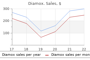
Cheap diamox 250mg without a prescription
The levator equipment is at risk in the inferior portion of the wound close to medications you cant donate blood buy 250 mg diamox otc the upper border of the tarsal plate symptoms 2 days before period purchase diamox 250mg otc, the place the levator fuses with the orbital septum. Excising muscle or opening of the septum is most secure if one stays away from the lower portion of the wound after excision of pores and skin. Persistent ptosis should be corrected by reinsertion of the levator into the tarsus or dermis. Lubrication (if needed-artificial tears for daytime dryness; Lacri-Lube ointment for nighttime protection) three. Wound care: the incision is gently cleaned with a cotton swab soaked in hydrogen peroxide three to four times daily, and ophthalmic antibiotic ointment is then utilized. Preoperative, A, and postoperative, B, views of a patient who had an upper eyelid blepharoplasty and a lower eyelid skin-muscle flap blepharoplasty. Extraocular muscle damage: the inferior oblique muscle lies between the medial and central adipose tissue compartments of the decrease lid. Care have to be taken to keep away from clamp, cautery, or sharp damage to this weak muscle throughout removing of adipose tissue. Persistent postoperative diplopia lasting longer than 1 to 2 weeks must be evaluated by an oculoplastic surgeon. Lower lid malposition: Excess "scleral show" within the quick postoperative period may be because of edema and transient paresis of the orbicularis oculi muscle. Tape support of the decrease lid can reduce the degree of scleral present, lower tearing, and improve consolation. Unappreciated preoperative decrease lid laxity may also contribute to postoperative ectropion. If the ectropion is due to overaggressive skin resection, correction will doubtless require complete scar release and full-thickness pores and skin grafting after several months of observation and conservative management. A transconjunctival strategy avoids the contraction associated with therapeutic of the pores and skin and muscle incisions used for the infraciliary strategy. In addition, patients with any diploma of laxity who require skin excision through an infraciliary approach must be thought-about for prophylactic lateral canthopexy. Retrobulbar hematoma/visual loss: this is the most feared complication of blepharoplasty and develops on account of vascular damage with retraction of the bleeding vessel into the retrobulbar house. Increasing intraocular stress can result in ophthalmoplegia, ischemia of the optic nerve, and visual loss. A retrobulbar hematoma typically develops throughout the first 4 to 6 hours postoperatively. Initial treatment entails simultaneous opening of incisions; iced saline compresses; 20% mannitol (2 g/kg) as an osmotic agent to decrease intraocular strain; acetazolamide (Diamox) 500 mg intravenously; dexamethasone (Decadron) 10 mg intravenously; management of hypertension (if present); head elevation; and correction of any coagulopathies. Any deterioration in visual acuity coupled with elevated intraocular pressure signifies impending optic nerve ischemia. In this situation, removal of all exterior sutures and lateral canthotomy/ inferior cantholysis are indicated on an emergency foundation for orbital decompression. In a 10-year retrospective evaluation of 892 patients present process blepharoplasty by a single surgeon, a self-reported dry eye questionnaire was administered to patients before and (minimum three weeks, imply follow-up 26 months) after surgical procedure. This highlights the need for all sufferers present process blepharoplasty to be assessed preoperatively for risks including scleral present, lagophthalmos, dryness, positive snap check end result (lower lid laxity), prior surgical procedure, and adverse vector, all of which may further enhance risk. Between the punctum for the lacrimal duct and medial adipose tissue pad of the decrease eyelid. Is greatest made via skin and the underlying orbicularis to facilitate flap elevation in a submuscular plane c. Management of retrobulbar hemorrhage would possibly embrace (circle all that may apply) a. Possible causes of scleral show/lid malposition include the entire following except a. Scarring and retraction between the anterior and middle lamella of the lower eyelid c. It permits for each functional and beauty issues to be addressed, resulting in above common patient satisfaction. One of the earliest signs of getting older is forehead ptosis coupled with an accumulation of redundant tissues in the glabella region and brow rhytids. A mixture of sun exposure and time induced facial pores and skin laxity allow brow tissue to migrate down previous the superior orbital rim creating a "bunching" of glabella and eyelid anatomy. Patients frequently current with useful complaints of visible impairment; the usually describe a weight or heaviness of their eyelids that limits their ability to stay attentive or focus as a result of impairment of vision and irritation of their eyelids weighing on their upper eyelid lashes. Brow shape and position have to be weighed against different variables corresponding to affected person acceptance, visibility of the scar, and risk of complications. Approaches to forehead shaping, elevating, and stabilizing have modified an excellent deal over the past several many years with newer advances allowing for a lower in visible scars, therapeutic time, and complications. Often a brow carry is coupled with blepharoplasty (staged or synchronous) for optimal practical and beauty benefit. Performing the brow raise previous to a blepharoplasty is really helpful to allow fixation of the brow position earlier than figuring out if the blepharoplasty is even necessary. If eyelid surgical procedure is performed, initial proper forehead stabilization allows precise measurements for eyelid skin markings. Personal or household history of bleeding issues or straightforward bruising is necessary to observe. Surgery: Previous surgical procedures of the higher face together with excision of pores and skin most cancers (and restore methods used to shut the defect), blepharoplasty, brow surgery, or traumatic accidents to the scalp/forehead are important to explore. Any history of postoperative nausea and vomiting ought to be explored in anticipation of preemptive perioperative pharmacologic intervention. Family historical past: Information on familial hairline recession and hair loss patterns in each female and male patients is important to think about when choosing the right brow lift strategy. Medication: the use of antiplatelet medicines, alcohol, or natural products Incidental: In anticipation of postoperative questions regarding swelling, routine inquiry as to the sufferers preferred sleeping position is needed. Preoperative dialogue of the affect of sleep place on dependent edema can get rid of concerns when sufferers expertise important asymmetry of eyelid or forehead swelling/bruising. Hairline place: the height of the brow is a vital variable in the selection of the proper forehead raise approach. Each method influences the hairline position and brow peak in unique ways as described later. Scalp: Hair thickness, colour, and high quality must be assessed to decide how visible a scar is likely to be when positioned around or behind the hairline. Eyelids: the presence of eyelid hooding as a outcome of dermatochalasis or eyelid ptosis ought to be noted. Facial nerve: Baseline facial nerve operate must be evaluated for strength and symmetry. Trigeminal nerve: Preoperative perform of the trigeminal nerve is important to notice as the supraorbital and supratrochlear branches could also be at risk through the process. Before (A, C) and after (B, D) pictures demonstrating typical results from a midforehead forehead lift strategy utilized in men. Other brow raise techniques are sometimes not as a lot an option for males with excessive or no hairline to mask the incision websites.
Purchase 250 mg diamox overnight delivery
Creating unnatural contours of the antihelix by overtightening the Mustard� sutures or leaving behind small fragments of cartilage after rasping 3 medications dictionary cheap diamox 250 mg amex. Overcorrection of lobule and superior side of auricle -reverse telephone deformity 5 medicine 8 discogs diamox 250 mg buy generic online. A much less conspicuous head-wrap or headband could also be worn thereafter for two to three weeks to keep ear position throughout initial healing. Pain medicine: Patients should be expected to have gentle ache after this operation. Pain beyond expectation within the early postoperative period could indicate a hematoma or early an infection. If utilizing a replaceable head-wrap, corresponding to a Glascock dressing, topical antibiotic ointment could additionally be use on the incision for three days followed by Aquaphor ointment, thereafter. Seroma or hematoma-may require needle aspiration or opening of incision for drainage 2. Wound infection and/or breakdown-Any purulence should be cultured and appropriate antibiotic management prescribed. A portion of the wound might must be opened to enable for drainage and healing by secondary intention. Surgical steps, together with excision of postauricular skin in fusiform trend, A, placement of short-term contouring sutures, B, and placement of Mustard� horizontal mattress sutures from behind the ear, C and D. External auditory canal meatal stenosis-Again, prevention is key by checking external auditory canal patency at the conclusion of the procedure and by placing conchomastoid sutures in a radial trend. Editorial Comment Otoplasty may be difficult techniquely for the surgeon however gratifying for the affected person. Knowledge of the normal anatomic contours of the ear is essential for the success of otoplasty. Like many facial plastic surgery procedures, a variety of described methods exist to accomplish the identical aim. The most important variations are for marking the antihelix, for cartilage contouring, and for cartilage excision. In my apply, I mark the specified suture places for the antihelix with needles positioned on the anterior facet of the pinna. Additionally, I tend to avoid any cartilage scoring or cartilage excision, if possible. Over the years, I have begun to slightly overcorrect the auricular deformity with the expectation that, over time, the ears tend to lateralize. Patients must be pre-operatively counseled that the vary of normal is type of broad with regard to the dimensions, shape, and projection of the pinna. The technique described above supplies a sound method to attaining the desired patient outcomes and creating a natural, reproducible look and symmetry between the ears. Modification of the Mustard� otoplasty technique utilizing momentary contouring sutures. There is little evidence to help or to oppose the use of antibiotic prophylaxis routinely for otoplasty. Of the 20 studies between 2000 and 2007 that met the inclusion criteria for evaluation, just one talked about a wound infection, with a price of three. Of the studies with no reported wound an infection, only 4 explicitly described use of prophylactic antibiotics. Thus, although proof is limited to help its use, I do use cephalexin by mouth for 7 days following the procedure, along with bacitracin ointment on the incision for three days. The most common deformities addressed in otoplasty for prominent ears embody all of the following, besides a. Nonsurgical correction of congenital auricular deformities within the early neonate: a preliminary report. Incisionless otoplasty: a dependable and replicable method for the correction of prominauris. This report discusses the postoperative outcomes of seventy two patients who underwent incisionless otoplasty. Hematology tests depend the number of white and red blood cells and platelets, measure the time necessary for blood to clot, and the aptitude of blood to carry oxygen throughout the body. The practitioner can also use hematology exams to determine whether or not the patient has inflammation or an infection. Hematology laboratory tests are also used to assess two pancreatic endocrine hormones secreted by the islet cells within the pancreas. When blood glucose is elevated, the pancreas secretes insulin, which causes glucose to cross the cell membrane allowing it to be used for energy, resulting in a decrease in blood glucose. When blood glucose ranges are low, the pancreas secretes glucagon, which signals the liver to release saved glucose into the blood, resulting in a rise in blood glucose. Blood glucose should be maintained within a slender vary, which happens naturally with the secretion of insulin and glucagon. However, failure of islet cells to properly function, caused by diseases similar to diabetes, can end result in excessive levels of blood glucose (hyperglycemia) or low ranges of blood glucose (hypoglycemia). The glomerulus in the kidneys acts as a filter to remove waste from the blood, which is collected in a renal tubule as urine. Metabolic waste corresponding to sodium, potassium, and phosphorus may be reused by the physique and is returned to the blood by the kidneys. This can occur suddenly (acute renal failure) in response to illness, medicines, accidents, and poisons. It can even occur slowly (chronic kidney disease) from diseases such as diabetes and hypertension. Chronic kidney disease can lead to end-stage renal illness, where all or almost all of the renal capabilities are permanently destroyed. Likewise, a person with one healthy kidney and one kidney with complete renal failure is said to have 50% renal operate. A person will expertise health issues if he/she has 25% or much less renal perform. Lipids are compounds used to retailer vitality, to develop cell membranes, and are components of nutritional vitamins and hormones. Cholesterol is released into the bloodstream mostly by the liver and other organs, though some ldl cholesterol is ingested in food. You will also find out about glucose exams and checks used to decide renal operate and lipid metabolism in this chapter. There is often a low stage of amylase in blood except the salivary glands or pancreas is blocked or broken. The amylase check can also be performed with the creatinine take a look at to help the healthcare supplier diagnose pancreatitis.
Diamox 250 mg generic with amex
Nursing Implications � Assess if the patient � Is undergoing radiation therapy � Is pregnant � Has had a blood transfusion in the earlier week � Has undergone a prostate biopsy in the past eight weeks � Is taking Bactrim treatment junctional rhythm best diamox 250mg, Septra medicine journey diamox 250mg discount mastercard, corticotrophin, Imuran, levodopa, Chloromycetin, methotrexate, or Cosmegen Understanding the Results � the reticulocyte take a look at outcomes can be found in 1 day. Vitamin B12 attaches to the intrinsic issue produced by the parietal cells in the abdomen. The intrinsic issue protects vitamin B12 from intestinal bacteria and permits absorption of vitamin B12 by the intestines. Up to 25% of the ingested vitamin B12 will usually be detected within the 24-hour urine sample. Rationale for the Test � Screen for � Absorption of vitamin B12 � Production of the intrinsic factor Nursing Implications � Assess if the patient � Is pregnant � Properly collected the 24-hour urine sample � Is taking Mycitracin, Dilantin, or colchicine � Has kidney disease � Used a laxative prior to administration of the check � Has had a radioactive scan 2 weeks previous to the take a look at Understanding the Results � the Schilling test outcomes can be found rapidly. Rouleaux settles quicker than erythrocytes, subsequently the elevated sedimentation rate signifies that the patient has irritation. Serum Osmolality Serum osmolality is the number of particles of gear which might be dissolved in blood serum (liquid portion of blood). A decrease in fluid results in an increase in serum osmolality-there is much less fluid within the blood. An improve in fluid results in a decrease in serum osmolality-there is extra fluid in the blood. The results of the urine check is in contrast with the serum osmolality to estimate kidney perform. Nursing Implications � Assess if the affected person � Has ingested alcohol, since this could affect the take a look at results � Recently acquired a blood transfusion Understanding the Results � Test outcomes can be found in 4 hours. Total Serum Protein the total serum protein take a look at assesses the levels of albumin, globulin, and total protein in a blood sample. It is constantly metabolized into amino acids, that are used to make enzymes, hormones, and new proteins. Albumin can additionally be essential for tissue progress and healing because it carries water, sodium, potassium, calcium, hormones, fatty acids; bilirubin, thyroxine and medicine to tissues. As a end result extra protein is on the market to the physique and blood can freely distribute globulins throughout the physique. Beta globulin binds with iron and transports iron all through the physique and is concerned in concentrating on international material so the foreign materials could be destructed by the immune system. A check for complete serum protein stories separate values for complete protein, albumin, and globulin. A toxicology test sometimes identifies if the drug is present but not the extent of the drug. Uric acid is produced when purine present in wine, beer, liver, anchovies, mackerel, and dried beans is metabolized. Uric acid is excreted by the kidneys by way of the urine and a small quantity in stool. Summary Hematology clinical laboratory checks study blood and its elements to assess for indicators of illness. The medical laboratory then measures the pattern and compares the results to a range of values that the medical laboratory has determined to be regular. A high stage of blood glucose signals the pancreas to secrete insulin, which assists glucose to cross cell membranes where glucose is used for power. Glucagon is a hormone that alerts the liver, muscular tissues, and other organs to launch saved glucose into the blood, leading to a rise in blood glucose ranges. The balance between insulin and glucagon secretions maintains a slender range of blood glucose. Estimate the sugar content material of the breakfast and subtract that quantity from the take a look at results. C-reactive protein is leaked from broken cells shortly after cells are injured growing the amount of C-reactive protein in blood. A low stage of C-reactive protein indicates that the liver is responding to scale back the irritation course of. C-reactive protein attaches to broken cells or microorganism enhancing phagocytosis within the destruction of the microorganism or broken cell shortly after cells are injured. C-reactive protein attaches to the liver inflicting a rise of white blood cells in blood to enhance phagocytosis within the destruction of the microorganism or broken cell shortly after cells are injured. What could be suspected if the patient has improve leukocyte rely but no signs of an infection Reschedule the check and inform the affected person to refrain from taking Prilosec 12 hour before coming for the take a look at. The concentration of electrolytes inside the body is on constant change and the stability is maintained by the kidneys. Cells within the kidney monitor the amount of sodium, potassium, and fluid in the bloodstream. Hormones from the kidneys (renin), adrenal gland (aldosterone), and the pituitary gland (antidiuretic hormone) are used to modify the electrolytes balance. These are calcium (Ca), magnesium (Mg), phosphate [contained in phosphorus (P)], potassium (K), sodium (Na), and chloride (Cl). Electrolyte tests are referred to as an electrolyte panel, primary metabolic panel, or complete metabolic panel. Basic metabolic and comprehensive metabolic panels measure electrolytes and different elements. Calcium Calcium (Ca) is required for growth of bone and teeth and for muscle contraction and blood clotting. As calcium will increase in blood, phosphate decreases and when phosphate increases in blood, calcium decreases. Too much calcium within the blood causes the thyroid gland to release calcitonin, which strikes calcium from blood to bone. There are two sorts of calcium blood exams which are performed as a part of a routine blood screening. Rationale for the Test � Screen for � Parathyroid gland perform � Kidney function � Kidney stones � Pancreatitis � Bone illness � Underlying trigger: � Muscle spasms, depression, confusion, tingling around the mouth and fingers, and muscle cramping and twitching are brought on by low calcium level in blood. Nursing Implications � Assess if the patient has � Taken milk, antacid, calcium salt, or calcium dietary supplements 8 hours earlier than administering the take a look at, since this can affect the check end result � Eaten or drunk 12 hours earlier than the check is administered � Taken Diamox, estrogen, albuterol, corticosteroids, vitamin D, lithium, laxatives, aspirin, theophylline, or contraception pills, since these medications can have an result on the take a look at result � Has had a blood transfusion just lately Understanding the Results � Test results can be found rapidly. If calcium levels in blood are abnormal, the healthcare supplier might order extra tests for the parathyroid hormone, vitamin D, alkaline phosphatase, acid phosphatase, and chloride, which can affect the calcium ranges in blood. Magnesium Magnesium (Mg), found principally in bones and inside cells, is an electrolyte and is used to switch potassium and sodium in and out of cells and to activate nerves, muscle tissue, and enzymes. The magnesium blood check measures the level of magnesium in a blood sample and is tested along with different electrolytes. Phosphate Phosphorus (P) is a mineral that accommodates a particle known as phosphate, which is critical for development of bones and tooth and for contracting muscle tissue.
Buy 250mg diamox with amex
Microcephaly with frank brain destruction and widespread encephalomalacia are widespread (12-11A) treatment 2 diamox 250mg cheap with mastercard. With few exceptions (toxoplasmosis and syphilis) medications for rheumatoid arthritis diamox 250 mg cheap without prescription, most congenital/perinatal infections are viral and are often secondary to transplacental passage of the infectious agent. The location and distribution of the calcifications might strongly counsel the particular infectious agent. Congenital syphilis is relatively rare, inflicting basilar meningitis, arterial strokes, and scattered dystrophic calcifications. Congenital, Acquired Pyogenic, and Acquired Viral Infections Pathology the timing of the gestational an infection determines the magnitude of brain insult. White matter volume loss occurs at all gestational ages and can be diffuse or multifocal. Microscopic examination reveals cytomegaly with viral inclusions in the nuclei and cytoplasm. Patchy and focal mobile necrosis, significantly of germinal matrix cells, is typical of first-trimester infection. Symptomatic newborns and infants might exhibit microcephaly, jaundice, hepatosplenomegaly, chorioretinitis, and rash. Sensorineural hearing loss, seizures, and developmental delay are the most important long-term risks. Infection, Inflammation, and Demyelinating Diseases 334 Newborns with systemic manifestations. The overwhelming majority of those newborns that show microcephaly, ventriculomegaly, cortical malformations. As a basic rule, the earlier the fetal an infection, the more extreme the findings (12-1) (12-4). Calcifications are predominantly periventricular, with a predilection for the germinal matrix zones, particularly the caudostriatal areas (12-2A). Calcifications range from numerous bilateral thick calcifications to faint punctate unilateral foci (122A) (12-3A) (12-4A). Cortical abnormalities such as cerebellar and hippocampal dysgenesis are well depicted (12-3C) (12-3D). Also, subependymal hyperintense foci of T1 shortening attributable to the periventricular calcifications may be seen. White matter hypointensities correspond to areas of demyelination and dysplasia. The pretemporal white matter cysts often begin as areas of T1 and T2 prolongation (12-4C) (12-5C). Cranial sonography is beneficial for evaluation of the neonatal and infant brain (up to 6-8 months of age). Other findings embody germinal zone cysts (germinolytic), which may be present alongside the caudostriatal grooves within the periventricular zones and within the anterior temporal white matter. Microcephaly and cortical dysplasia are also considerably much less widespread in congenital toxoplasmosis. The linear "tram-track" calcification sample described in some cases is nicely demonstrated right here. The an infection in humans is normally acquired from the ingestion of contaminated water or undercooked meals merchandise (usually fresh fruit, greens, and meat) or by direct contact with the feces of an infected cat. Ependymitis resulting in aqueductal obstruction and hydrocephalus with resultant macrocephaly is seen in approximately 50% of congenital toxoplasmosis. A diffuse inflammation of the meninges is present with large and small granulomatous lesions. Congenital toxoplasmosis causes severe chorioretinitis, jaundice, hepatosplenomegaly, development retardation, and brain injury. Infants with subclinical infection at delivery are in danger for seizures, in addition to delayed cognitive, motor, and visual defects. Note ribbon-like T2 shortening within the cortex reflecting hemorrhage and or Ca++. Early modifications include meningoencephalitis with necrosis, hemorrhage, and microglial proliferation. Hemorrhages may current as multifocal punctate, patchy, and curvilinear areas of hyperattenuation in the basal ganglia, white matter, and cortex (12-7A). In the acute and subacute levels of this disease, multifocal lesions (67%), deep grey matter involvement (58%), hemorrhage (66%), "watershed" pattern of damage (40%), and the occasional involvement of the brainstem and cerebellum have been reported. Areas of restricted diffusion may be the solely positive imaging findings in early cases. Late-stage illness shows severe volume loss with enlarged ventricles and multicystic encephalomalacia (12-9) (12-10). Hemorrhagic foci are common (66%) within 1 week of clinical analysis and finest detected with T2* sequences. Prevalence is higher in African Americans, low-income moms, and moms with multiple sexual partners. The risk is increased with main maternal infection in the course of the third trimester and could be decreased by cesarean supply. This disseminated an infection presents with lethargy, poor feeding, jaundice, hepatomegaly, seizures, and respiratory distress. Half of surviving infants have permanent deafness, vision loss, cerebral palsy, and/or epilepsy. Foci of patchy enhancement, sometimes a meningeal sample of enhancement, are common on T1 C+ scans. Zika virus has been instantly linked to severe fetal microcephaly in infants born to contaminated moms. The virus leads to neurotoxicity and in experimental models impaired human neurosphere growth. Clinical Issues the diagnosis of Zika virus an infection in the grownup is complicated by the truth that up to 80% of infected individuals are asymptomatic. Conjunctivitis and Guillain-Barr� syndrome are uncommon medical manifestations of the infection. The affected new child exhibits microcephaly, a nonspecific time period that refers to a smaller than anticipated head for regular gestational age. In Zika virus infection and other congenital infections, insults to the developing brain result in microencephaly (small brain), which leads to a small head (microcephaly). Also, associated overlapping sutures, closed fontanelles, and redundant scalp skin folds may be clinically noticed. Seizures, poor feeding, hypotonia, and lethargy are nonspecific common medical options among severely affected newborns. The virus is generally transmitted by contaminated feminine mosquito Imaging Cerebral parenchymal calcifications are universally current. Cerebral, cerebellar, and mind stem volume loss, ventriculomegaly, and resultant microencephaly are seen. Other reported abnormalities include occipital periventricular cysts, demyelination, microphthalmia, and cataracts. High charges of chorioretinitis and hydrocephalus are noticed, thus often mimicking the imaging options of toxoplasmosis.

Diseases
- Sener syndrome
- Cerebellar degeneration, subacute
- Common cold
- Glaucoma, congenital
- Blastomycosis
- Pipecolic acidemia
- Chromosome 22, trisomy q11 q13
- Warts
- Gastrocutaneous syndrome
- Phosphoglycerate kinase 1 deficiency
250 mg diamox buy amex
To accomplish this medications via g-tube diamox 250mg order amex, the now free-floating part of hyoid bone is grasped with an Allis or Leahy clamp and pulled anteroinferiorly treatment sinus infection buy diamox 250mg low price, and a cylinder of tissue at the base of the tongue is created by way of a superiorly aimed dissection with an insulated needle-tipped cautery. Exposure may be facilitated by using the first suture placed as a traction suture and cutting it after later sutures have been positioned. The tongue is grasped with a perforating towel clip, and an assistant pulls the tongue anteriorly. The surgeon locations a long 18-Ga spinal needle through the pores and skin overlying the body of the hyoid bone and advances the tip through the tongue mucosa on the foramen cecum. The stylet is removed and a #2 braided polyester suture is advanced through the needle, the end is grasped within the mouth with a tonsil clamp, and the needle is withdrawn. The traction suture is divided and the intraoral part removed by the anesthesiologist. The pharyngotomy is rigorously closed in layers with interrupted absorbable suture over a Penrose drain. Pressure on the retractor brings the base of the tongue into the wound, facilitating the excision of the block of tissue at the foramen cecum. Dermal attachments or irritation could require removal of a fusiform part of overlying pores and skin. The cyst is elevated and a aircraft is developed posterior to the physique of the hyoid bone. B, the cyst is recognized, the strap muscles are retracted, and the cyst is pedicled on the hyoid bone. The vallecula is entered posterior to the hyoid bone: Only a thin layer of tissue may separate the mucosa from the posterior floor of the hyoid. The dissection might stray from the midline: Landmarks have to be checked incessantly. Too little tongue tissue could also be removed: � Surgeons have reported low recurrence charges with elimination of solely a really restricted cuff of tissue above the body of the hyoid. The strap muscular tissues have to be introduced together medially to help fill the lifeless house created by the loss of the midportion of the hyoid bone and the superficial delicate tissues. Routine pain administration with acetaminophen, ibuprofen, and oxycodone could also be began immediately. Placement of a traction suture to guide the tongue-base resection by way of the foramen cecum. Use of a traction suture to ship the higher part of the tongue base into the wound to improve publicity of the higher a part of the resection. When untreated, recurrent infections, fistulas to skin, and indicators of obstruction from growing size of the lesion can occur. The Sistrunk procedure is a comparatively safe process with low issues and dangers, and when carried out properly, this has a really low recurrence risk for the cyst. However, there are risks related such as inflammation, hematoma, fever, or hypersensitivity. A transoral strategy, endoscopically assisted, has been reported just lately with the benefit of not producing an exterior scar. Risk of recurrence in kids operated for thyroglossal duct cysts: a systematic review. Preoperative computed tomography of suspected thyroglossal duct cysts in children underneath 10 years of age. Recurrent thyroglossal duct cysts: a 23-year expertise and a new technique for management. Recurrence: Incomplete excision of secretory material, nearly invariably located in the tongue base if the hyoid has been beforehand resected, might produce a pseudocyst situated virtually anyplace within the anterior neck. Treatment requires reexcision by way of a method such as the suture-guided transhyoid pharyngotomy or other strategy to the tongue base. Primary surgical excision using the Sistrunk procedure is the beneficial management. Thyroglossal duct cysts in adults treated by ethanol sclerotherapy: a pilot research of a nonsurgical method. Such anomalies are second only to thyroglossal duct cysts as the commonest plenty of congenital origin and are the most common congenital plenty presenting in the lateral neck. The differential prognosis of branchial cysts contains dermoid cysts and lymphatic vascular malformations. The physical presence of a cutaneous sinus or fistula tract strengthens this analysis. The surgical management of these anomalies varies depending on the cleft or pouch of origin and whether the anomaly is a cyst, sinus, or fistula. A detailed knowledge of the embryology of those lesions is subsequently essential for each definitive diagnosis and correct therapeutic intervention. Many of the symmetrically paired structures of the top and neck come up from the branchial system, which appears in the course of the fourth to seventh weeks of fetal development as six ridges on the superolateral floor of the pinnacle. These ridges are known as branchial arches owing to their resemblance to the gill arches of fish. Each arch represents a condensation of mesoderm, from which cartilage, muscle, and bone will type. The arches are separated from each other by an external cleft of ectodermal origin and an internal pouch of endodermal origin. As fetal maturation proceeds, many of the exterior clefts and internal pouches resorb and the arches fuse. Anomalies of the branchial system can form from any aberration on this course of together with failure of the external clefts and inner pouches to recede. Anomalies of the branchial system current in any of three distinct types: sinus tracts, fistula tracts, and cysts. A fistula tract has each cutaneous and pharyngeal communications and is assumed to come up from a persistence of each cleft and pouch with dissolution of the dividing plate. A branchial cyst is basically a residual pouch or cleft with no external or internal communication. Branchial anomalies mostly arise from the second (65% to 90%) and first (8% to 25%) branchial techniques; anomalies of the third and fourth systems are comparatively rare. Branchial cleft anomalies with cutaneous sinus or fistula tracts are usually obvious at start; cysts and branchial pouch anomalies with pharyngeal communications are inclined to present later in life. Within the grownup inhabitants, the imply age of diagnosis of branchial cysts has been reported as 40 years, with a slight female predominance. First Branchial Anomalies First branchial cleft anomalies are often divided into two subgroups or types. The cutaneous cervicofacial opening, if present, is typically at or below the angle of the mandible and at all times superior to the level of the hyoid. The course of the sinus or fistula tract or location of the cyst is very variable with respect to the parotid gland and facial nerve. Tracts could additionally be superficial or deep to the main branches of the facial nerve and may even course between particular branches. Third Branchial Anomalies Anomalies of the third branchial system are most frequently sinus tracts or cysts; the prevalence of a real fistula is very rare.
Diamox 250mg buy lowest price
In this case medicine man dr dre diamox 250mg buy without prescription, the healthcare provider is prone to medicine reaction diamox 250mg generic with amex perform a cervical cerclage, which quickly closes the cervix until the mother enters labor. If the birth is premature, the healthcare supplier could perform a cranial ultrasound to decide if there were issues brought on by the premature start. The new child is often administered the sweat test that helps determine if the newborn has a high stage of chloride in his sweat, which may be a sign of cystic fibrosis. About the sixteenth week of gestation, the healthcare provider may perform amniocentesis, which is the removing of some amniotic fluid. Fetal cells include the amniotic fluid which is analyzed to decide if the fetus has a delivery defect. These embody cystic fibrosis, Duchenne muscular dystrophy, sickle cell anemia, thalassemia, hemophilia, and Tay-Sachs disease. Amniocentesis is also performed to decide if the fetus is Rh-positive when the mom has the Rh issue. She should also call her healthcare supplier if she experiences fever, ache, or cramping in her stomach. If the affected person is unable to fill her bladder, a urinary catheter is inserted into the urethra and saline might be infused into the bladder. The healthcare provider will order completely different tests to further assess the fetus and mom. A girl who carries this mutated gene and has a household historical past of breast or ovarian cancer could have a better than regular chance of creating these cancers. If so, some sufferers might resolve to have a mastectomy and/or oophorectomy to forestall these cancers from growing. Patients who test optimistic can also be advised to take tamoxifen to inhibit this gene. Breast Ultrasound A breast ultrasound is used to examine suspicious findings of a mammogram because ultrasound expertise can differentiate between a cyst and strong tissue. Furthermore, the breast ultrasound can examine areas of the breast that are tough to view on a mammogram. Cervical Cerclage (Weak Cervix) If the patient has an incompetent cervix, the cervix would possibly open previous to the thirty seventh week of gestation and will result in untimely delivery. The healthcare supplier could perform a cervical cerclage, which is a procedure to shut the cervix, to make sure that the cervix remain closed till after the thirty seventh week of gestation. Rationale for the Test � To prevent premature opening of the cervix Nursing Implications � Determine if the patient � Has signed a consent type � Has any allergies � Has been taking anticoagulant medication such as Plavix, Coumadin, heparin, or aspirin � Can lie on her again � Is able to lie nonetheless � Has been treated for infection of the pelvis, cervix, or vagina � Has not eaten or drunk for 12 hours before the process � Does not have uterine contractions � Does not have vaginal bleeding � Does not have ruptured membranes Understanding the Results � the process takes lower than 1 hour. Chorionic Villus Sampling the placenta contains chorionic villi, which are tiny growths that contain the identical genetic material because the fetus. Chorionic villus sampling is a process during which a sampling of chorionic villi is biopsied between the tenth and twelfth week of gestation and is examined to decide if the fetus has a genetic disorder. A name must be made if she experiences fever, swelling on the insertion site or is dizzy. The contraction stress take a look at determines if the fetus will remain wholesome during natural childbirth. However, some fetuses can become negatively affected by the decrease oxygen level, so the healthcare provider would possibly determine a caesarean start. During the contraction stress check, a fetal coronary heart monitor is attached to the mom while she is administered oxytocin. The fetal coronary heart fee is anticipated to decelerate throughout a contraction and accelerate following the contraction. The dose is elevated till there are three contractions, every lasting more than 45 seconds over a period of 10 minutes. Rationale for the Test � Assess � Ability of the fetus to remain healthy throughout natural childbirth � Health of the placenta Nursing Implications � Determine if the affected person � Has signed a consent type � Has any allergies � Has been taking anticoagulant medicine corresponding to Plavix, Coumadin, heparin, or aspirin � Can lie on her back � Is in a position to lie nonetheless � Is capable of follow instructions � Has eaten or drunk 8 hours earlier than the check � Has smoked 2 hours before the check � Has emptied her bladder Understanding the Results � the procedure takes lower than 2 hours. Cordocentesis If amniocentesis or other checks reveal that the fetus might need anemia, the healthcare provider might order a cordocentesis to confirm the finding, usually within the second trimester. A cordocentesis is the sampling of fetal blood from the umbilical cord to determine if the fetus has a blood dysfunction or is Rh-positive. She must also call her healthcare provider if she experience fever, ache, or cramping in her stomach. Cranial Ultrasound A cranial ultrasound is carried out on premature newborns to assess complications which may have arisen in the course of the premature birth. Rationale for the Test � To screen for galactosemia Nursing Implications � Assess if the affected person � Has had a blood transfusion � Has reduction from galactosemia. Karyotyping Karyotyping is a take a look at that determines the number and quality of chromosomes in a cell and is used to detect potential genetic issues. Tissue samples for karyotyping are typically taken throughout chorionic villus sampling or amniocentesis. Rationale for the Test � To assess for fetal genetic dysfunction Nursing Implications � Determine if the affected person has undergone genetic counseling Understanding the Results � the procedure takes less than 1 hour. Pap Smear A Pap smear is a procedure that removes sample cells from the cervix to assess if there are any abnormal cells. Further examination is necessary if the sample is positive, indicating irregular cells on the cervix. If so, the patient might not really feel snug having a pelvic examination or a Pap smear carried out. Sperm Penetration Tests Sperm penetration tests are carried out, when a woman is having problem changing into pregnant, to decide if the sperm can transfer through the cervical mucus and into the fallopian tubes. The two kinds of sperm penetration checks are � Sperm penetration assay: this test mixes sperm with hamster eggs to see if the sperm can penetrate the egg. Sweat Test the sweat check is run to a newborn between the ages of two days and 5 months to assess if the new child might need cystic fibrosis. Rationale for the Test � To assess for cystic fibrosis Nursing Implications � Determine if the patient � Is lower than four weeks of age. Vaginosis Tests Vaginosis is irritation of the vulva and vagina attributable to an infection or a response to an irritant, leading to a painful vaginal discharge and itching. The commonest causes of vaginosis are � Candida albicans: it is a yeast an infection that causes lumpy white discharge and itching. The 4 vaginosis exams are � Whiff test: Potassium hydroxide answer is dropped on the sample. Only the yeast stays on the slide, indicating that the affected person has a yeast infection. The laboratory technician then identifies, by way of microscopic examination, the organism causing the infection. If so, the affected person might not feel snug having a sample of the discharge taken. Any suspicious area of the breast or cervix is examined carefully by taking a tissue sample or a biopsy of an abnormal growth to determine it.
250 mg diamox buy fast delivery
Carbon monoxide can end result in immediate death Carbon monoxide is a colorless medicine z pack cheap diamox 250mg with mastercard, odorless gas Carbon monoxide attaches to hemoglobin changing oxygen Carbon monoxide is irreversible 245 medicine 9 minutes diamox 250 mg order on-line. The patient was recently contaminated with hepatitis A virus the patient at some point was infected with hepatitis A virus the patient was just lately contaminated with hepatitis B virus the affected person sooner or later was infected with hepatitis B virus 246. Bacteria in the gut break down protein the heme causes the liver to create ammonia the heme causes the kidneys to create ammonia Ammonia is the outcomes of breakdown of hemoglobin 248. Why would strenuous train earlier than the cardiac enzyme research affect the check outcomes Enzymes being studied are contained in other muscle cells that may be injured during exercising B. Accumulation of fatty acid in the mind and nerve cells No accumulation of fatty acid within the mind and nerve cells the presence of HexosaminidaseA enzyme in the new born Accumulation of protien in the mind and nerve cells 253. The sensitivity take a look at identifies the microorganism the sensitivity take a look at identifies treatment that kills the microorganism the sensitivity take a look at identifies erythrocytes the sensitivity check identifies leucocytes 254. A false-positive end result if the H pylori depend is low and undetectable Results are never conclusive A false-negative outcome if the H pylori rely is low and undetectable the check will kill the microorganism 256. To assess temporomandibular joints To assess sinuses To assess jaw All of the above 258. An amphetamine Adderall Dexedrine A sedative Medical TesTs and Procedures deMysTified 603 259. To assess the absorption of vitamin B12 Screen for being pregnant To assess the vitamin B6 degree To assess the vitamin folic acid level 261. It is a handheld meter that helps determine if the new child has jaundice It measures the extent of bilirubin by being positioned on the skin It measures the extent of bilirubin in a newborn All of the above 262. Administer a sedative per order Cancel the check Wait for a calmer second to administer the check Tell the affected person to behave like an adult 263. Assess the velocity, path, and move of blood Assess fluid content material of the brain Assess changes within the brain chemistry All of the above 264. A radioactive tracer being injected into a blood vessel Contrast material being injected into a blood vessel the patient inhaling a fuel None of the above 265. Pure-tone audiometry Speech reception/Word recognition Whispered speech check Otoacoustic emissions test 267. To assess the underlying cause of hyperthyroidism To assess the underlying reason for hypothyroidism To assess the remedy of hypopituitarism All of the above 268. Order a 24-hour urine free cortisol test Diagnose the patient with Cushing syndrome Diagnose the affected person with hyperthyroidism Diagnose the affected person with uncontrolled diabetes 271. Converts vitamin D to an active form Increases absorption of calcium by the intestine Causes the kidneys to retain calcium All of the above 272. What does the practitioner do with the afP check outcomes if the ultrasound gestation age is totally different from the estimated gestation age Adjust the check results using the multiple of median (MoM) issue Medical TesTs and Procedures deMysTified 605 C. Call her practitioner Call the radiologist Ask the patient to lie down Call for emergency medical assist immediately 275. What would you do if the patient feels short of breath during a transvaginal ultrasound Reposition the affected person or elevate the head of the bed Stop the take a look at Tell the affected person to take deep breaths Distract the patient by asking the patient about her youngsters 277. What different test would possibly the practitioner orders together with the cardiac enzymes check Why ought to a affected person not take tranquilizers 5 days before the electronystagmogram (enG) check Tranquilizers might gradual eye motion giving a false check end result Tranquilizers might enhance eye motion giving a false test end result Tranquilizers would possibly trigger nausea and vomiting during the test Tranquilizers would possibly cause nausea and vomiting following the take a look at Pure-tone audiometry Speech reception/word recognition Whispered speech test Otoacoustic emissions check Amsler grid test Perimetry take a look at Tangent display check Snellen test Amsler grid take a look at Perimetry take a look at Tangent display check Snellen test 283. What may the practitioner do if the patient is identified with cardiac tamponade To assess blood circulate through the guts To assess blood circulate by way of veins To assess blood circulate through the mind To assess blood move via the eyes 290. What may a rise in bilirubin levels from an amniocentesis taken after the twentieth week indicate Be refrigerated Be stored in a gallon container Be discarded All of the above 295. Using the thyroid gland scan Using the salivary gland scan Using the liver scan Using the bladder scan 296. What is your response if a affected person tells you that he/she spit into the sterile container when you asked for a sputum culture C-reactive protein is leaked from damaged cells shortly after cells are injured increasing the amount of C-reactive protein in blood B. A low stage of C-reactive protein signifies that the liver is responding to reduce the inflammation process C. C-reactive protein attaches to damaged cells or microorganism enhancing phagocytosis in the destruction of the microorganism or damaged cell shortly after cells are injured D. C-reactive protein attaches to the liver inflicting a rise of white blood cells in blood to improve phagocytosis within the destruction of the microorganism or damaged cell shortly after cells are injured Medical TesTs and Procedures deMysTified 609 Medical Tests and Procedures Demystified Answers 1. Tracer materials helps map the route that cancer cells spread from the most cancers website by way of the lymphatic system. To assess liver harm as a result of unintended or intentionally overdosing with acetaminophen. Coumadin decreases coagulation time, thereby rising the danger of bleeding in the course of the procedure. Screening for cancer, assessing cancer treatment, and assessing for the success of surgery to remove the tumor 24. Chorionic villus sampling could be performed earlier within the pregnancy than amniocentesis. Avoid eating and ingesting aside from water 12 hours before the check is run. A sputum cytology research cells contained in the sputum and a sputum culture identifies microorganism within the sputum. Assess if the affected person carried out strenuous train before the test was administered. Maintains voltage across cell membranes and carry electrical impulses within the physique B. Arrange for a sign that the patient can provide to the practitioner to indicate that the patient is uncomfortable during the procedure. A common lung scan by which a ventilation scan is carried out after which a perfusion scan is carried out 147. This check identifies heterophil antibodies that kind between 2 and 9 weeks after the affected person becomes contaminated.
Diamox 250 mg purchase on line
Mechanical thrombectomy can also be suitable in sufferers beyond the therapeutic window or in whom thrombolytic therapy is contraindicated stroke treatment 60 minutes discount diamox 250 mg visa. The primary targets of emergent stroke imaging are (1) to distinguish "bland" or ischemic stroke from intracranial hemorrhage and (2) to select/triage patients for potential reperfusion therapies medications to treat bipolar disorder 250 mg diamox. Once intracranial hemorrhage is excluded, the second important concern is determining whether or not a significant cerebral vessel is occluded. Nontraumatic Hemorrhage and Vascular Lesions 212 choice for depicting doubtlessly treatable major vessel occlusions. With helical acquisition, the whole protocol could be completed inside 15 minutes as a single examination with separate contrast boluses. The most particular however least delicate signal is a hyperattenuating vessel crammed with acute thrombus. It is critically important to establish calcified cerebral emboli, as they carry a close to 50% danger of repeat ischemic stroke. M1-3 represent the middle cerebral artery cortex with every space allotted one point. The insular cortex (I), lentiform nuclei (L), caudate head (C), and inner capsule are scored with one point each. Loss of the insular cortex ("insular ribbon" sign) (8-38A) and decreased density of the basal ganglia ("disappearing basal ganglia" sign) are the commonest findings (8-37A). Cortical gyriform enhancement is uncommon in early arterial occlusion however may occur in late acute/early subacute infarction. Nontraumatic Hemorrhage and Vascular Lesions 214 occlusion with a "retrievable" intravascular thrombus present Note hypodensity of the proper temporal lobe, insular cortex ("insular ribbon signal"). The core infarct includes the proper frontal lobe, basal ganglia, and deep/periventricular white matter. Arterial Anatomy and Strokes the standard color scale is graduated from shades of pink and yellow to blue and violet. Well-perfused grey matter appears red/yellow, white matter appears blue, and ischemic brain is blue/purple. Here the colour scales are reversed to emphasize the abnormally extended transit time within the ischemic mind. The infarct core is seen as a darkish blue/purple or black space that contrasts with the normally perfused red/yellow mind (837B). Ischemia-induced vascular injury predisposes to two highly morbid and potentially deadly postischemic issues, i. With massive vessel occlusions, loss of the anticipated "flow void" in the affected artery can generally be recognized. Also look carefully for the presence of multifocal parenchymal microbleeds in older patients. In this age group, "blooming black dots" are mostly attributable to persistent hypertension or amyloid angiopathy. The presence of cerebral microbleeds could also be an impartial danger factor for subsequent anticoagulation-related hemorrhage. Aquaporins are transmembrane proteins-water channels-that facilitate bidirectional selective water transport out and in of the cell. Arterial Anatomy and Strokes to intraarterial thrombolysis or mechanical thrombectomy. Clot location and size may be precisely determined and collateral circulation delineated. Frequent findings embody an abrupt vessel "cut-off" (8-42A), "meniscus" signal, tapered or "rat-tail" narrowing, or "tram-track" appearance with a trickle of contrast across the intraluminal thrombus. Other widespread angiographic findings embrace a "naked" or "bare" area of nonperfused mind (8-42B) (8-42C), gradual antegrade filling with delayed washout of distal branches (seen as intraarterial contrast persisting into the capillary or venous phase), and pial collaterals with retrograde filling across the cortical watershed (8-42D) (8-42E) (8-42F). Less widespread signs are hyperemia with a vascular "blush" across the infarcted zone (so-called luxurious perfusion) (8-42F) and "early draining" veins (arteriovenous shunting with distinction showing in veins draining the infarct while the rest of the circulation remains to be within the late arterial or early capillary phase). Mass effect is uncommon in hyperacute stroke however quite common in the acute/late acute phases. A "hyperdense vessel" sign could be simulated by elevated hematocrit (all the vessels seem dense, not simply the arteries), arterial wall microcalcifications, and hypodense mind parenchyma. Nontraumatic Hemorrhage and Vascular Lesions 218 (8-41A) Acute stroke in a 47y man reveals patchy hyperintensity in left caudate nucleus, lateral putamen, and parietal cortex. Arterial Anatomy and Strokes Subacute Cerebral Infarcts Terminology Strokes evolve pathophysiologically with corresponding changes reflected on imaging studies. Frank tissue necrosis with progressive inflow of microglia and macrophages around vessels ensues with reactive astrocytosis around the perimeter of the stroke. Ischemiadamaged vascular endothelium turns into "leaky," and blood-brain barrier permeability will increase. When reperfusion is established-either spontaneously or following therapy with tissue plasminogen activator-exudation of red blood cells by way of the damaged blood vessel walls causes parenchymal hemorrhages. Petechial hemorrhages are more widespread than lobar bleeds and are most common in the basal ganglia and cortex. Mass impact initially increases, then begins to lower by 7-10 days following stroke onset. Patchy or gyriform enhancement appears as early as 2 days after stroke onset, peaks at 2 weeks, and generally disappears by 2 months. Signal depth in subacute stroke varies depending on (1) time since ictus and (2) the presence or absence of hemorrhagic transformation. Signal intensity decreases with time, reaching isointensity at 1-2 weeks (the T2 "fogging effect") (8-47). Nontraumatic Hemorrhage and Vascular Lesions 222 can generally be identified as a well-delineated hyperintense band that extends inferiorly from the infarcted cortex alongside the corticospinal tract. The intravascular enhancement often seen within the first 48 hours following thromboembolic occlusion disappears within three or four days and is changed by leptomeningeal enhancement brought on by persisting pial collateral blood move. Patchy or gyriform parenchymal enhancement can happen as early as 2 or three days after infarction (8-46) and should persist for 2-3 months, in some circumstances mimicking neoplasm (8-48). Arterial Anatomy and Strokes Chronic Cerebral Infarcts Terminology Chronic cerebral infarcts are the tip result of ischemic territorial strokes and are also called postinfarction encephalomalacia. A cavitated, encephalomalacic brain with strands of residual glial tissue and traversing blood vessels is the standard gross look of an old infarct (8-49A). The adjacent sulci and ipsilateral ventricle enlarge secondary to quantity loss within the affected hemisphere (849A). Look for atrophy of the contralateral cerebellum secondary to crossed cerebellar diaschisis.
