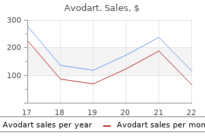
Avodart 0.5 mg buy without a prescription
Intralesional triamcinolone suspension injected or iontophoresed into the plaques and nodules has proven blended results symptoms gluten intolerance discount avodart 0.5 mg on line. Oral therapies include tocopherol (vitamin E) symptoms adhd buy avodart 0.5 mg, paraaminobenzoate, colchicine, tamoxifen, and acetyl-L-carnitine, but information supporting oral therapy are weak. Extracorporeal shock wave therapy may reduce penile ache but may worsen curvature. Knuckle pads are sometimes related to Dupuytren contracture, clubbing, or camptodactylia (irreducible flexion contracture of a number of fingers). Some instances are familial, and some are associated to trauma or frequent knuckle cracking. Knuckle pads have additionally been related to autosomal dominant epidermolytic palmoplantar keratoderma with a mutation in keratin 9. They are differentiated clinically from the nodular sort of neurodermatitis and from the small, hemispherical pitted papules that may develop over the knuckles after frostbite or in acrocyanosis, and from rheumatic nodules. DevataS,etal: Desmoid tumors: a complete review of the evolving biology, unpredictable habits, and myriad of management choices. Collagenousfibroma(desmoplasticfibroblastoma) this slow-growing, deep-set, benign fibrous tumor is usually positioned in the deep subcutis, fascia, aponeurosis, or skeletal muscle of the extremities, limb girdles, or head and neck regions. It is characterized by hypocellularity and dense bands of hyalinized collagen that may infiltrate into skeletal muscle. Despite this, no tumors have been reported to metastasize or recur after excision. Chromosomal translocation (2; 11)(q31; q12) in addition to trisomy eight have been reported. NishioJ,etal: Translocation t(2;11) is characteristic of collagenous fibroma (desmoplastic fibroblastoma). Aponeuroticfibroma Aponeurotic fibroma has additionally been known as juvenile aponeurotic fibroma (calcifying fibroma). It is a tumorlike proliferation characterised by the appearance of slow-growing, cystlike lots that happen on the limbs, especially the hands and ft. Histologically, the distinctive lesions are sharply demarcated and composed of collagenous stroma showing acid mucopolysaccharides infiltrated by plump mesenchymal cells with oval nuclei. Pachydermodactyly Pachydermodactyly represents a benign fibromatosis of the fingers. There is a fullness of the medial and lateral digit just proximal to the proximal interphalangeal joint. This asymptomatic course of most often is first famous in adolescence and usually entails multiple fingers. Five types have been described: traditional, localized, transgrediens (abnormality extends to metacarpophalangeal areas), familial, and pachydermodactyly related to tuberous sclerosis. Patients with pachydermodactyly associated with repetitive tics respond to therapy for the obsessive-compulsive dysfunction. Infantilemyofibromatosis Infantile myofibromatosis is the commonest fibrous tumor of infancy. Eighty percent of patients have solitary lesions, with half of these occurring on the pinnacle and neck. Congenital generalized fibromatosis is an unusual situation that presents at birth or quickly after. Skeletal lesions, primarily of the metaphyseal regions of the lengthy bones, happen in 50% of sufferers. If solely the skin and bones develop fibromas, the prognosis is superb, with spontaneous decision of the lesions and with no complications anticipated in the first 1�2 years of life. Desmoidtumor Desmoid tumors occur as massive, deep-seated, wellcircumscribed plenty arising from the muscular aponeurosis. They most frequently happen on the belly wall, especially in ladies during or quickly after pregnancy. Desmoid tumors have been divided into five varieties: abdominal wall, extraabdominal, intra-abdominal, a number of, and those occurring in Gardner syndrome/familial adenomatous polyposis. They recur domestically and might kill in the event that they invade, surround, or compress very important constructions. The most harmful desmoid tumors are subsequently these on the root of the neck and the intra-abdominal type. Treatment may be with wide local excision, radiotherapy, or hormonal manipulation. Mesenteric desmoid tumors have been handled with 600 Diffuseinfantilefibromatosis this course of occurs throughout the first 3 years of life and is normally confined to the muscular tissues of the arms, neck, and shoulder space. There is multicentric infiltration of muscle fibers with fibroblasts resembling those in aponeurotic fibromas. Infantile fibromatosis may be seen in any location, although the arms, legs, and trunk are the same old websites. Nodular tumors of the scalp, face, and extremities usually seem in early childhood. Histologically, fibroblasts with nice, intracytoplasmic eosinophilic granules are embedded in a homogeneous eosinophilic dermal floor substance. Ultrastructurally, the fibroblasts demonstrate faulty synthesis of collagen, deposited as fibrillogranular material. DenadaiR,etal: Systemic hyalinosis: new terminology, severity grading system, and surgical approach. Histologically, the dermis is regular, but the dermis is infiltrated with proliferating myofibroblasts and collagen bundles. Eosinophilic cytoplasmic inclusions in many of the fibroblasts are attribute. Treatment by surgical excision has a excessive risk of recurrence, and conservative, nonsurgical administration is commonly acceptable. Spontaneous regression is mostly noted, but the lesion might cause functional impairment and may infiltrate deeply before regression occurs. Mohs micrographic surgery has been carried out successfully using each trichrome staining and smooth muscle actin staining to reveal the inclusion our bodies inside tumor cells. SpingardiO,etal: Infantile digital fibromatosis: our expertise and long-term results. Overlying pores and skin changes are unusual however could embody increased hair, alteration in pigmentation, and eccrine gland hyperplasia with hyperhidrosis. Most instances are solitary, but multiple tumors have been reported; 91% of lesions are famous throughout the first year of life, and 23% are congenital. Most lesions occur in the axillary region, upper arm, upper trunk, inguinal region, and external genital space. Biopsy reveals an organoid pattern with several varieties of tissue organized in whorls or bands.
Reishi Mushroom. Avodart.
- How does Reishi Mushroom work?
- Boosting the immune system, high blood pressure, high cholesterol, viral infections, tumors, prostate cancer, inflammatory diseases, heart disease, asthma, bronchitis, stress, kidney disorders, liver disease, HIV disease, altitude sickness, fatigue, chronic fatigue syndrome (CFS), insomnia, stomach ulcers, poisoning, herpes-related pain, shingles, and other conditions.
- Are there safety concerns?
- Are there any interactions with medications?
- What is Reishi Mushroom?
Source: http://www.rxlist.com/script/main/art.asp?articlekey=96871
0.5 mg avodart discount amex
Mucosal lesions are also seen in half of sufferers with bilateral conjunctival congestion medications prescribed for migraines 0.5 mg avodart generic overnight delivery, aphthouslike oral lesions symptoms 14 days after iui 0.5 mg avodart discount fast delivery, gingivitis, glossitis, and with extension down the throat, dysphagia. The oral lesions can have a gray exudate or small (tapioca granule) white lesions on the taste bud. In this stage, supportive care can maintain the patient until the spontaneous eradication of the virus. Neutralizing antibodies from survivors and experimental antivirals are being examined as therapies. In this group, the viral ailments of dermatologic interest are measles (rubeola) and German measles (rubella). Other viruses of this group are mumps virus, parainfluenza virus, Newcastle illness virus, and respiratory syncytial virus. Cases of measles continue to be imported into the United States, which have resulted in quite a few "outbreaks" due to important numbers of nonimmune individuals. Also generally recognized as rubeola and morbilli, measles was a worldwide illness that the majority typically affected children beneath 15 months of age. In the current epidemics, nonetheless, older youngsters, adolescents, and adults may be affected. Measles is spread by respiratory droplets and has an incubation period of 9�12 days. The prodrome consists of fever, malaise, conjunctivitis, and outstanding higher respiratory signs (nasal congestion, sneezing, coryza, cough). After 1�7 days, the exanthem seems, normally as macular or morbilliform lesions on the anterior scalp line and behind the ears. Lesions are most prominent and confluent in the initially involved areas and may be more discrete on the extremities. The spots seem first on the buccal mucosa nearest to the lower molars as 1-mm white papules on an erythematous base. Highly effective two-dose vaccines are available, and when international locations attain a fee of 95% vaccination, measles elimination has been achieved. However, measles remains a major well being downside in many countries, including developed countries that present immunizations to their population. Numerous hospitalizations and even deaths from measles are still occurring in these developed nations. The majority of cases are in unvaccinated individuals, supporting the idea that vaccination (specifically two doses) is protecting, and that these measles epidemics and deaths are preventable. Low vaccination levels exist in these countries for many reasons, each philosophic and socioeconomic. In an Australian outbreak, these methods prevented 80% of attainable secondary circumstances. Rubella Rubella, generally generally recognized as German measles, is brought on by a togavirus and doubtless spreads by respiratory secretions. There is a prodrome of 1�5 days consisting of fever, malaise, sore throat, eye ache, headache, red eyes, runny nose, and adenopathy. The exanthem begins on the face and progresses caudad, covering the whole physique in 24 h and resolving by the third day. The lesions are usually pale-pink, morbilliform macules, smaller than these of rubeola. Posterior cervical, suboccipital, and postauricular lymphadenitis happens in additional than half of instances. Arthritis and arthralgias are common complications, especially in adult girls, lasting 1 month or longer. The analysis is confirmed by discovering rubella-specific IgM in oral fluids or the serum. This IgM develops rapidly, however 50% of sera drawn on the primary day of the rash are negative. However, the virus is present in oral secretions for 5�7 days after the rash has appeared. Modified measles happens in a partially immune host on account of prior infection, persistent maternal antibodies, or immunization, and this is a milder disease. The course is shorter, the exanthem less confluent, and Koplik spots may be absent. A analysis of measles is established by the presence of a excessive fever, Koplik spots, the characteristic conjunctivitis, upper respiratory symptoms, and typical exanthem. Biopsies of skin lesions may present syncytial keratinocytic large cells, just like those seen in respiratory secretions. Identification of virus-specific IgM (5 days after the rash presents) is extremely suggestive of an infection in an unimmunized individual. If done too early, however, a serum IgM assay could lead to a falsenegative end result, and the take a look at must be repeated. The mixture of IgM serologic testing and virus isolation is the current gold standard for prognosis. Rubella, scarlet fever, secondary syphilis, enterovirus infections, and drug eruptions are within the differential prognosis. Administration of excessive doses of vitamin A will scale back the morbidity and mortality of hospitalized children with measles. Live virus vaccination is recommended at age 12 months, with a booster before entering faculty (age 4�5 years). Prophylaxis with vaccination and immune globulin ought to be supplied to uncovered susceptible individuals. It should be supplied inside the first few days after publicity, so identification of susceptible individuals is critical. Numerous different manifestations, such as glaucoma, microcephaly, and varied visceral abnormalities, may emerge. Among the cutaneous expressions are thrombocytopenic purpura; hyperpigmentation of the navel, brow, and cheeks; bluish purple, infiltrated, 2�8 mm lesions ("blueberry muffin" type), which represent dermal erythropoiesis; persistent urticaria; and reticulated erythema of the face and extremities. CaserisM,etal: French 2010�2011 measles outbreak in adults: report from a Parisian educating hospital. GiustiD,etal: Virological analysis and management of two circumstances of congenital measles. SheikineY,etal: Histopathology of measles exanthem: a case with characteristic options and eosinophils. It happens in youngsters eight months to 10 years of age, however most circumstances are between 2 and 3 years. The cause is unknown, however a viral origin has been proposed as a end result of it occurs in young children and is seasonal, and secondary circumstances in families have been reported. No reproducible viral etiology has been implicated; however, at least three circumstances attributed to parvovirus B19 have been reported. The lesions are often discrete, 1-mm erythematous papules that coalesce to poorly marginated morbilliform plaques. Lesions begin unilaterally near a flexural space, usually the axilla (75% of cases). Spread is centrifugal, with new lesions showing on the adjoining trunk and proximal extremity.
0.5 mg avodart purchase mastercard
Urinary values larger Bonus pictures for this chapter may be found on-line at expertconsult medicine journal impact factor buy avodart 0.5 mg on line. The term "epidermal nevus" includes a quantity of entities shinee symptoms mp3 order avodart 0.5 mg with amex, including keratinocytic epidermal nevi, nevus sebaceus, and nevus comedonicus, relying on which epidermal cell or construction comprises the lesion. The epidermal nevus should be classified by its predominant histologic and scientific function: keratinocytic, comedonal, or sebaceous. This suggests that all "epidermal nevi" should be categorized based on their histologic phenotype. Epidermal nevi of every kind are thought of an expression of cutaneous mosaicism with genetic mutation in the affected skin, however sparing the unaffected skin in widespread lesions; a lot much less regularly, the mutation is discovered not only in the pores and skin, but additionally in other tissues. Lesions follow the lines of Blaschko, suggesting that they symbolize postzygotic mutations. In general, larger lesions, extra widespread lesions, and lesions of the pinnacle and neck usually tend to have associated inner issues. The combination of an epidermal nevus and an related internal downside is known as "epidermal nevus syndrome. Keratinocyticepidermalnevi Keratinizing epidermal nevi are the most typical kind of epidermal nevus and are described by an excellent variety of phrases, similar to linear epidermal nevus, hard nevus of Unna, soft epidermal nevus, and nevus verrucosus (verrucous nevus). If the lesion is widespread on half the physique, the term nevus unius lateris has been used. The most typical pattern of keratinocytic epidermal nevus is linear epidermal nevus. The individual lesions are verrucous, skin-colored, dirty-gray, or brown papules, which coalesce to type a serpiginous plaque. Interspersed in the localized patch could also be attractive excrescences and barely comedones. The age of onset of epidermal nevi is generally at delivery, but they might also develop throughout the first 10 years of life. The histologic modifications within the dermis are hyperplastic and have an effect on mainly the stratum corneum and stratum malpighii. It is assumed that every of those varieties can be related to a particular mutation in the affected skin that, if widespread, would give rise to the cutaneous dysfunction with the identical histology. For example, epidermal nevi that present epidermolytic hyperkeratosis would have the same gene mutation because the disorder of cornification, bullous congenital ichthyosiform erythroderma. In reality, sufferers with this kind of epidermal nevus might have gonadal mosaicism that can lead to offspring with the full-blown disorder. These identical gene mutations are found in sporadic seborrheic keratoses, which, not surprisingly, have the identical histology. Various abnormalities of the bones, vessels, and mind are associated with these clinical findings. Large keratinocytic epidermal nevi of the trunk and extremities are more regularly related to skeletal abnormalities. Since each nevus sebaceus and keratinocytic epidermal nevi had been included within the original and huge reports of epidermal nevus syndrome, the exact characterization of the "keraticocytic epidermal nevus syndrome" stays to be outlined. Epidermal hyperplasia, with acanthosis, papillomatosis, parakeratosis, and hyperkeratosis, is also present (the options of a keratinocytic epidermal nevus). In rare instances, as a substitute of half the physique being affected, large quadrants of the physique, favoring folds, are the websites of the epidermal growths (ptychotropism). Epidermal nevus syndrome may be related to vitamin D�resistant hypophosphatemic rickets, perhaps from circulating fibroblast progress issue 23 acting as a phosphaturic. Rarely, keratinocytic and adnexal malignancies occur in keratinocytic epidermal nevi. Any newly showing lesion inside a steady epidermal nevus should be biopsied to exclude this risk. Management of keratinocytic epidermal nevi is tough as a outcome of, except the remedy extends into the dermis (and thus might cause scarring), the lesion recurs. AkhyaniM,etal: Bullous ichthyosiform erythroderma in a child born to a parent with systematized linear epidermolytic hyperkeratosis. MehraS,etal: A novel somatic mutation of the 3-hydroxysteroid dehydrogenase gene in sporadic cutaneous verruciform xanthoma. PanagiotopoulosA,etal: Assessment of cryotherapy for the remedy of verrucous epidermal naevi. ParadelaS,etal: Epidermal nevi treated by carbon dioxide laser vaporization: a collection of 25 patients. Nevuscomedonicus Nevus comedonicus is characterised by carefully organized, grouped, typically linear papules that have at their center dilated follicular openings with keratinous plugs resembling comedones. Cysts, abscesses, fistulas, and scars develop in about half the instances, which have been described as "inflammatory" nevus comedonicus. As with other epidermal nevi, lesions could additionally be localized to a small space or could have an intensive distribution. The lesions may develop any time from start to age 15 however are usually present by age 10. Follicular tumors, including trichofolliculoma and pilar sheath acanthoma, can appear within the lesion. Histologic examination reveals massive dilated follicles full of orthokeratotic attractive material and lined by atrophic squamous epithelium. The interfollicular dermis is papillomatous, as seen in typical epidermal nevi. Treatment of lesions not sophisticated by inflammatory cysts and nodules is primarily beauty. Pore-removing beauty strips and comedone expression might enhance the beauty appearance. Keratinocytic nevi are seen in at least 4 epidermal nevus syndromes, as follows: 1. A linear soft, thick, papillomatous keratinocytic nevus within the absence of cerebriform hyperplasia of the palms and soles, however with segmental glomerulosclerosis. Fibroblast growth factor receptor 3 epidermal nevus syndrome (Garcia-Hafner-Happle syndrome). A velvetytype nonepidermolytic epidermal nevus and cerebral defects identify this syndrome. Sebaceous nevus coexists with aplasia cutis, normally in close proximity to each other. Systematized, linear, velvety, orthokeratotic nevus with hypertrichosis and follicular hyperkeratosis. Diffuse ichthyosis-like hyperkeratosis covers the whole body, including the palms and soles. Each variant has attribute cutaneous findings and at occasions relatively specific internal findings.
Buy avodart 0.5 mg low cost
Relatedpalisadinggranulomas Interstitial granulomatous dermatitis with arthritis is a situation with a range of clinical shows medications used for depression 0.5 mg avodart buy with amex. It can present with spherical to oval erythematous or violaceous plaques on the flanks treatment herniated disc avodart 0.5 mg generic mastercard, axillae, inside thighs, and decrease stomach. Severe systemic vasculitis inflicting renal disease has responded to corticosteroids with or without cyclophosphamide. Mycophenolate sodium and rituximab have both been used to deal with extreme manifestations related to Sj�gren syndrome. Arthritis might happen before, during, or after the eruption and tends to affect a number of joints of the higher extremities. Some displays are paraneoplastic, associated with a range of stable and hematopoietic tumors. Histologically, a average to dense inflammatory infiltrate is seen via the reticular dermis, composed mostly of histiocytes distributed interstitially around discrete bundles of sclerotic collagen. The eruption is usually asymptomatic and may spontaneously involute after many months or years. If remedy is required, intralesional corticosteroids, methotrexate, etanercept, ustekinumab, tocilizumab, and cyclosporine have been used. It usually presents with eroded or ulcerated, symmetrically distributed umbilicated papules or nodules on the elbows, knuckles, and knees. The biopsy could reveal leukocytoclastic vasculitis and collagen degeneration in early lesions or palisaded granulomatous infiltrates with dermatofibrosis and scant neutrophilic particles in older lesions. Methotrexate-induced papular eruption seems in patients with rheumatic ailments throughout methotrexate therapy. They present with erythematous indurated papules, usually situated on the proximal extremities. Histopathologic examination reveals an inflammatory infiltrate composed of histiocytes interstitially organized between collagen bundles of the dermis, intermingled with few neutrophils. At occasions, small rosettes composed of clusters of histiocytes surrounding a thick, central collagen bundle are current within the deep reticular dermis. An eruption consisting of evanescent, nonpruritic, salmon-pink, macular, or papular lesions on the trunk and extremities might precede the onset of joint manifestations by many months. The systemic symptoms of fever and serositis usually recur over weeks every afternoon. The dose-response curve for methotrexate plateaus with parenteral administration of 15 mg/ m2/week. Refractory disease has been handled with pulse methylprednisolone, tocilizumab, and cyclophosphamide. Biopsy demonstrates a fibroblastic proliferation with a collagenous stroma various from clean muscle actin� positive cellular fascicles to paucicellular areas with randomly organized spindle or stellate cells. Standard therapy consists of immunosuppressive agents, typically methotrexate and oral corticosteroids, although some patients have responded to physical remedy with out immunosuppressive therapy. Patients with new onset of rheumatologic illness ought to be screened for indicators and symptoms suggesting neoplasm. Symmetricsynovitis Symmetric seronegative synovitis is an idiopathic type of arthritis sometimes related to idiopathic edema. Both genders are equally affected, with age at onset often within the fourth to fifth decade. During bouts of irritation, the bright-red involvement of the ears is confined to the cartilaginous portion while the earlobes stay conspicuously regular. There may be conductive deafness because of the obstruction produced by the swollen cartilage. The nasal septal cartilage is equally involved to produce rhinitis, with crusting and bleeding and finally saddle nose. Involvement of the bronchi, larynx, and epiglottis produces hoarseness, coughing, and dyspnea. Patients evaluated for chest pain are often released without therapy and with a diagnosis of costochondritis. Cell-mediated immunity to cartilage has been demonstrated in vitro, with a level of response correlated with disease exercise. A second connective tissue illness or other autoimmune illness is current in about one third of sufferers with relapsing polychondritis, and a few cases seem to be paraneoplastic, occurring in affiliation with hematopoietic malignancies. Histologically, a predominantly neutrophilic infiltrate is noted in the perichondrium. Dapsone, 100 mg a few times daily for an adult, reduces the frequency of flares but is often inadequate to control relapsing polychondritis. Sustained response to etanercept has been reported, even after failure to reply to infliximab. Endobronchial ultrasonography has been used to facilitate the analysis of relapsing polychondritis and estimate the scale of the involved airway for placement of stents. These acid mucopolysaccharides, produced by fibroblasts, are extremely hygroscopic, binding about a thousand times their very own volume in water. They are important in holding water in the dermis and are responsible for dermal volume and texture. Normally, the sulfated acid mucopolysaccharide chondroitin sulfate and heparin are the primary dermal mucins. In sure ailments, fibroblasts produce abnormally large quantities of acid mucopolysaccharides, usually hyaluronic acid. These acid mucopolysaccharides (mucin) accumulate in large quantities in the dermis and may be visible as pale-blue, granular or amorphous materials between collagen bundles. They are often not visualized with hematoxylin and eosin stains as a end result of the water they bind is removed in processing, so the presence of increased mucin is suspected by the presence of huge, empty areas between the collagen bundles. Acid mucopolysaccharides can be detected by special stains, similar to colloidal iron, alcian blue, and toluidine blue. Incubation of the tissue with hyaluronidase eliminates the staining, confirming the presence of hyaluronic acid. Increased dermal mucin might end result from many illnesses and is a standard element of wound therapeutic. The mucinoses are illnesses by which manufacturing of increased amounts of mucin is the first process. The genetic ailments by which mucin accumulates as a outcome of inherited metabolic abnormalities are termed the mucopolysaccharidoses (see Chapter 26). The main lesions are a number of, waxy, 2�4 mm, domeshaped or flat-topped papules. The dorsal arms, face, elbows, and extensor extremities are most incessantly affected. On the glabella and forehead, coalescence of lesions leads to the prominent furrowing of a "leonine facies. Dysphagia from esophageal involvement usually occurs, and the abdomen or gut may be affected. Pulmonary problems with dyspnea brought on by restrictive or obstructive disease are also frequent.
Avodart 0.5 mg cheap line
In some patients medications used for anxiety avodart 0.5 mg cheap without prescription, an extensive patchy treatment lupus 0.5 mg avodart generic with mastercard, scaly eruption covers most of the trunk, buttocks, and extremities. Generally, tinea infection of the arms is of the dry, scaly, and erythematous type suggestive of T. Other areas are frequently affected at the same time, especially the mix of each ft and one hand. Interdigital tinea should also be distinguished from gram-negative toe web infection. Diabetic patients develop interdigital fungal infections at a youthful age than patients without diabetes. Trichophyton mentagrophytes usually produces acutely inflammatory multilocular bullae. The burning and itching that accompany the formation of the vesicles might cause nice discomfort, which is relieved by opening the tense vesicles. The fissures between the toes, in addition to the vesicles, may turn into secondarily infected with pyogenic cocci, which may result in 291 Tineaofhandsandfeet 15 Diseases Resulting from Fungi and Yeasts recurrent attacks of lymphangitis and inguinal adenitis. The sweat between the toes and on the soles has a high pH, and damp keratin is an effective culture medium for the fungi. Dermatophytid of the palms may be related to inflammatory tinea of the toes and begins with the appearance of teams of minute, clear vesicles on the palms and fingers. As a rule, both palms are involved, and the eruption tends to be symmetric; in some instances, nonetheless, just one hand is affected. Because the illness usually begins on the feet, the affected person ought to be suggested to dry the toes completely after bathing. The use of a great antiseptic powder on the feet after bathing, significantly between the toes, is strongly suggested for prone persons. Tolnaftate powder (Tinactin) or Zeasorb medicated powder is an excellent dusting powder for the toes. Plain talc, cornstarch, or rice powder could additionally be dusted into socks and footwear to maintain the feet dry. Periodic use of a topical antifungal agent may be required, especially when sizzling occlusive footwear is worn. Diagnosis Demonstration of the fungus by microscopic examination of the scrapings taken from the involved website establishes the analysis. Copious dry scale from the instep, heel, and sides of the foot could be gathered by scraping with the sting of a glass microscope slide. Bullae ought to be unroofed, and either the entire roof is mounted intact or scrapings are produced from the underside of the roof. The mycelia could also be seen beneath low-power microscopy, however higher observation of each hyphae and spores is obtained by the 10� goal with the condenser cranked down or the light aperture closed by two-thirds. The traces of juncture of normal epidermal cells dissolve into a branching network which will easily be mistaken for fungus buildings ("mosaic false hyphae"). Treatment Clotrimazole, miconazole, sulconazole, oxiconazole, ciclopirox, econazole, ketoconazole, naftifine, terbinafine, flutrimazol, bifonazole, efinaconazole, and butenafine are effective topical antifungal brokers. Aluminum chloride 10% solution or aluminum acetate, 1 part to 20 parts of water, may be useful. Topical antibiotic ointments, similar to gentamicin (Garamycin), which would possibly be efficient against gram-negative organisms are useful additions in some moist interdigital lesions. In the ulcerative kind of gram-negative toe internet infections, systemic antibiotic remedy is critical (see Chapter 14). Keratolytic brokers containing salicylic acid, resorcinol, lactic acid, and urea could also be helpful in some circumstances, though all might result in maceration if occluded. Treatment of fungal infection of the skin of the feet and palms with griseofulvin, 500�1000 mg/day, may be efficient. Recommended adult dosing for terbinafine is 250 mg/day for 1�2 weeks; for itraconazole, 200 mg twice every day for 1 week; and for fluconazole, 150 mg once weekly for 4 weeks. Abbreviated schedules and intermittent dosing with other brokers could also be potential but require further examine. In one small study, itraconazole, a hundred mg twice daily, was given instantly after meals on 2 consecutive days. Onychomycosis(tineaunguium) Onychomycosis is outlined because the infection of the nail plate by fungus and represents as a lot as 30% of diagnosed superficial fungal infections. Distal subungual onychomycosis primarily entails the distal nailbed and the hyponychium, with secondary involvement of the underside of the nail plate of fingernails and toenails. White superficial onychomycosis (leukonychia trichophytica) is an invasion of the toenail plate on the floor of the nail. Proximal subungual onychomycosis involves the nail plate mainly from the proximal nailfold, producing a particular clinical picture. Candida onychomycosis produces destruction of the nail and massive nail bed hyperkeratosis. White superficial onychomycosis is the name given to one sort of superficial nail an infection brought on by this fungus by which small, chalky white spots appear on or in the nail plate. Infection normally begins at the lateral edge of the nail, burrows beneath the plate, and produces massive portions of tacky debris. Nattrassia mangiferae (Hendersonula toru loidea) and Scytalidium hyalinum have been reported to cause onychomycosis, as properly as a moccasin-type tinea pedis. In addition to the extra common features of onychomycosis, similar to nail plate thickening, opacification, and onycholysis, options of infection with these fungi include lateral nail invasion alone, paronychia, and transverse fracture of the proximal nail plate. In a rural school in Mexico the place most people wear nonocclusive leather-based sandals, Trichosporon cutaneum, Candida spp. Cutaneous Scytalidium infections are widespread in sufferers from the tropics, especially the West Indies and Africa. They usually carry the organism with them, even once they emigrate to more temperate climates. A yellowish discoloration happens, which spreads proximally as a streak within the nail. Later, subungual hyperkeratosis turns into outstanding and spreads until the whole nail is affected. Gradually, the whole nail turns into brittle and separated from its mattress on account of the piling up of subungual keratin. Fingernails and toenails present a similar appearance, and the pores and skin of the soles is more probably to be concerned, with characteristic branny scaling and erythema. The an infection typically begins with scaling of the nail underneath the overhanging cuticle and remains localized to a portion of the demonstration of fungus is made by microscopic examination or by culture. Samples obtained by a drilling approach may have the next yield than these obtained by curettage. Culture on Sabouraud agar with chloramphenicol and cycloheximide (Mycosel) agar was 32% delicate. Both office and central laboratories can be used to isolate fungi, however false-negative outcomes are common in both settings.
Buy generic avodart 0.5 mg on line
JacoblsA symptoms 22 weeks pregnant purchase avodart 0.5 mg with mastercard,etal: Systemic evaluate of the rapidity of the onset of motion of topical remedies within the remedy of mild-to-moderate acne vulgaris medicine park oklahoma 0.5 mg avodart buy mastercard. Complications Even with the superb remedy choices available, scarring may occur. This could additionally be quite outstanding and infrequently outcomes from the cystic kind of acne, though smaller lesions may produce scarring in some individuals. Pitted scars, widemouthed depressions, and keloids, primarily seen alongside the jawline and chest, are widespread forms of scarring. ManolacheL,etal: A case of strong facial oedema efficiently treated with isotretinoin. MargolisD,etal: Potential association between the oral tetracycline class of antimicrobials used to deal with pimples and inflammatory bowel disease. MorroneA,etal: Clinical features of zits vulgaris in 444 sufferers with ethnic skin. This kind is characterised by quite a few comedones (many of which are double or triple) and enormous abscesses with interconnecting sinuses, cysts, and grouped inflammatory nodules. The cysts happen on the again, buttocks, chest, forehead, cheeks, anterior neck, and shoulders. Hidradenitis suppurativa and dissecting cellulitis of the scalp could also be seen with acne conglobata, an association known as the "follicular occlusion triad. Athletes and bodybuilders ought to be questioned about the usage of anabolic steroids, which can induce such aggressive pimples. Pretreatment with prednisone and low initial doses of isotretinoin, as described for acne fulminans, are beneficial to keep away from flaring of disease. BruzzeseV: Pyoderma gangrenosum, pimples conglobata, suppurative hidradenitis, and axial spondyloarthritis. It is characterized by highly inflammatory nodules and plaques that endure swift suppurative degeneration, leaving ragged ulcerations, totally on the chest and back. As in pimples conglobata, anabolic steroids taken by bodybuilders could induce this condition. Prednisone, 40�60 mg, is necessary in the course of the preliminary 4�8 weeks to calm the dramatic inflammatory response of zits fulminans. This ought to be slowly increased to normal doses and continued for a full 120�150 mg/kg cumulative course. Nodular, cystic, and pustular lesions happen mainly on the back, buttocks, and thighs. Acne tropicalis usually happens in younger adults who may have had pimples vulgaris at an earlier age. This is very true of these within the armed forces stationed in the tropics and carrying backpacks. Treatment is that for cystic zits, however pimples tropicalis might persist until the patient moves to a cooler, much less humid climate. Skin findings could embrace acne fulminans, pimples conglobata, pustular psoriasis, hidradenitis suppurativa, dissecting cellulitis of the scalp, Sweet syndrome, Sneddon-Wilkinson disease, and palmoplantar pustulosis. The chest wall and mandible are the most typical websites for musculoskeletal complaints in adults; the long bones, significantly the tibia, predominate in children. Bone modifications of the anterior chest wall on nuclear scans are probably the most particular diagnostic findings. If isotretinoin is used, it must be initiated at a low dosage, similar to 10 mg/day, together with prednisone for the primary month to forestall flaring of the disease. Anakinra, methotrexate, sulfasalazine, and cyclosporine are different, much less well-documented however doubtless efficient choices. Pamidronate and other bisphosphonates corresponding to ibandronate, alendronate, and zoledronic acid, are efficient in treating the osteoarticular manifestations. ColinaM,etal: Clinical and radiologic evolution of synovitis, pimples, pustulosis, hyperostosis, and osteitis syndrome. GaladariH,etal: Synovitis, acne, pustulosis, hyperostosis, and osteitis syndrome handled with a mixture of isotretinoin and pamidronate. Acneestivalis Also often identified as Mallorca zits, this rare type of zits starts within the spring, progresses in the course of the summer season, and resolves completely within the fall. Dull-red, dome-shaped, onerous, small papules, normally not larger than 3�4 mm, develop on the cheeks and normally lengthen on to the sides of the neck, chest, shoulders, and characteristically the higher arms. The primary lesions are trivial or even nonexistent, however the compulsive neurotic habit of choosing the face and squeezing minute comedones produces secondary lesions that crust and will leave scars. If the affected person admits to picking however being unable to stop this behavior, enchancment could. Breaks in the epithelium and spillage of follicular contents into the dermis lead to the lesions. If secondary to a drug, an eruption begins inside days of initiation of the treatment, could additionally be accompanied by fever and malaise, and resolves when the drug is stopped. Acneiform eruptions might originate from skin exposure to varied industrial chemical substances, similar to fumes generated in the manufacture of chlorine and its byproducts. These chlorinated hydrocarbons could trigger chloracne, consisting of cysts, pustules, folliculitis, and comedones. The most potent acneiforminducing brokers are the polyhalogenated hydrocarbons, notably dioxin (2,3,7,8-tetrachlorodibenzo-p-dioxin). Cutting and lubricating oils, pomades, crude coal tar utilized to the skin for medicinal purposes, heavy tar distillates, coal tar pitch, and asbestos are identified to trigger acneiform eruptions. Acneiform eruptions are induced by medications corresponding to iodides from radiopaque contrast media or potassium iodide, bromides in medication corresponding to propantheline bromide, testosterone, cyclosporine, antiepileptic medicines, lithium, and systemic corticosteroids. When medium or excessive doses of corticosteroids are taken for as briefly as 3�5 days, a particular eruption might occur, often known as steroid zits. It is a sudden outcropping of inflamed papules, most numerous on the upper trunk and arms. The lesions typically present as papules somewhat than comedones; however, a histologic examine confirmed they start follicularly with microcomedone formation. Topical steroids, especially the fluorinated types or when applied underneath occlusion, can also induce an acneiform eruption. Epidermal progress issue inhibitors, including monoclonal antibodies and tyrosine and multikinase inhibitors used in cancer therapy, produce a folliculitis within the majority of handled sufferers. Often, oral minocycline and topical benzoyl peroxide are given prophylactically on the outset of the cancer therapy to stop what could also be a dose-limiting reaction. Comedonal lesions could additionally be limited to the nasal crease, within the flexural areas in youngsters and on the temple and malar pores and skin in Favre-Racouchot syndrome. LiT,etal: Skin toxicities associated with epidermal development issue receptor inhibitors. During antibiotic therapy, patients develop both superficial pustules 3�6 mm in diameter, flaring out from the anterior nares, or fluctuant, deep-seated nodules. Culture of these lesions normally reveals a species of Klebsiella, Escherichia coli, Enterobacter, or from the deep cystic lesions, Proteus. With long-term, broad-spectrum antibiotic remedy, the anterior nares might turn into colonized with these gram-negative organisms. As the usage of long-term antibiotic remedy declines, this illness has turn out to be much less common.
Syndromes
- Abdominal ultrasound
- VLDL: 2 - 30 mg/dL
- What illnesses, diseases, and disorders are you being treated for?
- Breathing may be rapid
- Take the medicines your doctor told you to take with a small sip of water.
- Are there tiny red or purple spots on the skin (petechiae)?
Avodart 0.5 mg cheap on-line
These are essentially the most commonly prescribed medicines and are particularly helpful for the papulopustular patients and some patients with the erythrotelangiectatic sort medications in canada avodart 0.5 mg buy generic. Benzoyl peroxide and topical clindamycin medicine man lyrics order 0.5 mg avodart mastercard, alone or together, are sometimes beneficial and well tolerated by the glandular subset of rosacea patients. If oral antibiotics are wanted, the topical products could also be used to preserve remission after discontinuance of oral preparations. These drugs calm inflammation and abate symptoms but require temporary (no longer than 1 week) pretreatment with a potent topical corticosteroid to be tolerated initially. Many rosacea patients could tolerate a nighttime application of tretinoin if Cetaphil lotion is used instantly before use. Retinoids could help repair sun-damaged skin and normalize a variety of the abnormalities current. The -adrenergic receptor agonist brimonidine is out there as a gel for the treatment of facial redness. It is applied once within the morning, which induces vasoconstriction for up to 12 hours. HsuC-C,etal: Carvedilol for the remedy of refractory flushing and persistent erythema of rosacea. RallisE,KorfitisC: Isotretinoin for the remedy of granulomatous rosacea: case report and evaluate of the literature. TanghettiE,etal: Consensus recommendations from the American Rosacea Society on the administration of rosacea. Edema and at instances an intense reddish or cyanotic erythema accompany this pustular course of. It is distinguished from zits by the absence of comedones, fast onset, fulminating course, and absence of pimples on the back and chest. Pyoderma faciale is differentiated from rosacea by the inconsistent history of flushing, the absence of preexisting erythema or telangiectases of the convex portions of the face, and the big abscesses and nodules. This condition is also called rosacea fulminans, a designation many favor after Plewig categorized it as such. Oral steroids are given for a number of weeks, followed by the addition of isotretinoin, 10�20 mg, increasing to zero. Steroids might usually be discontinued after several weeks of isotretinoin, but the latter ought to be given for a full 120�150 mg/ kg whole dose. Because sufferers are predominately ladies of childbearing age, being pregnant points require full discussion. In such sufferers, amoxicillin, erythromycin, azithromycin, or clindamycin, all being pregnant category B medication, could also be thought-about. FuentelsazV,etal: Rosacea fulminans in being pregnant: successful treatment with azithromycin. It is a particular dermatitis confined symmetrically around the mouth, with a clear zone of about 5 mm between the vermilion border and the affected pores and skin. The use of fluorinated topical corticosteroids is probably the most regularly identified cause. Treatment of perioral dermatitis includes discontinuing topical corticosteroids or protecting the skin from the inhaled product. In patients without steroid exposure, oral or topical antibiotics and topical adapalene, azelaic acid, and metronidazole have all been profitable in clearing the eruption. Periorbitaldermatitis Periorbital (periocular) dermatitis is a variant of perioral dermatitis occurring on the decrease eyelids and skin adjacent to the upper and decrease eyelids. If intranasal inhaled corticosteroids are used, a perinasal distribution may be seen. SchwarzT,etal: A randomized, double-blind, vehicle-controlled study of 1% pimecrolimus cream in adult sufferers with perioral dermatitis. The discrete papules appear as yellowish brown lesions on diascopy and as caseating epithelioid cell granulomas histologically. Long-term remedy with minocycline or isotretinoin could also be used, typically with gratifying results. These different entities, which meet no other standards for rosacea other than having pink papules on the face, are included right here. Besides extremity and truncal lesions, several ladies had dramatic lesions of the labia majora. Children with skin of color (Afro-Caribbean, African American, and Asian) dominate the reports, but white patients are also prone. Topical metronidazole, erythromycin, sulfacetamide-sulfur mixtures, and an oral macrolide or tetracycline-type antibiotic all are sometimes effective. In some sufferers, the mix of prednisone and dapsone has proved helpful. Al-MutairiN: Nosology and therapeutic choices for lupus miliaris disseminatus faciei. AmiruddinD,etal: Clinical evaluation of 35 cases of lupus miliaris disseminatus faciei. GutteR,etal: Childhood granulomatous periorificial dermatitis in youngsters with extrafacial involvement. KoikeY,etal: Lupus miliaris disseminatus faciei efficiently treated with tranilast: report of two cases. These cutaneous signs may be an indication of a generalized systemic course of or just an isolated superficial occasion. Patients with atopic dermatitis and syndromes with atopic-like dermatitis are also predisposed to bacterial infections. The categorization of bacterial infections on this chapter first addresses diseases caused by gram-positive micro organism, followed by these attributable to gram-negative micro organism, after which several miscellaneous diseases attributable to the rickettsiae, mycoplasmas, chlamydiae, and spirochetes. PetryV,etal: Bacterial skin colonization and infections in sufferers with atopic dermatitis. However, bullae, widespread erythema and desquamation, or vegetating pyodermas may be indicators of Staphylococcus aureus an infection. Two distinctive cutaneous lesions that occur with endocarditis are the Osler node and Janeway lesion or spot. The Osler node is a painful, erythematous nodule with a pale center located on the fingertips. The Janeway spot is a nontender, angular hemorrhagic lesion of the soles and palms. Staphylococcus aureus is a normal inhabitant of the anterior nares in 20�40% of adults and in addition resides on the palms and perineum in smaller numbers of individuals. Spread of an infection within the hospital setting is incessantly traced to the arms of a well being care worker. Proper handwashing method is crucial in preventing this nosocomial complication.
Generic avodart 0.5 mg otc
It grows on most routine mycologic media medicine 3d printing avodart 0.5 mg cheap free shipping, however cycloheximide will suppress growth of Prototheca spp xanthine medications 0.5 mg avodart discount amex. The use of fluorescent antibody reagents permits the fast and reliable identification of Prototheca spp. Intravenous amphotericin B remains the best agent for disseminated Prototheca infections. Surgery, in addition to topical amphotericin B and doxycycline, has been used for isolated cutaneous illness. Pathogenic bacteria, particularly Pseudomo nas aeruginosa, are sometimes discovered concurrently. The colonization may be benign, but malignant otitis could often happen, especially in diabetic or iatrogenically immunosuppressed sufferers. Inoculation from an exogenous source (primary inoculation tuberculosis, tuberculosis verrucosa cutis) 2. Endogenous cutaneous unfold contiguously or by autoinoculation (scrofuloderma, tuberculosis cutis orificialis) three. Hematogenous unfold to the pores and skin (lupus vulgaris; acute miliary tuberculosis; tuberculosis ulcer, gumma, or abscess; tuberculous cellulitis) (Lupus vulgaris can also occur adjacent to lesions of scrofuloderma, suggesting that each hematogenous unfold and local spread are able to triggering this response pattern. Tuberculous ulcer/abscess/cellulitis and tuberculosis cutis orificialis are circumstances of poor host immunity against Mycobacterium tuberculosis. As host immunity develops, the skin test becomes optimistic, and the variety of organisms on biopsy diminishes. This is partly attributable to the persistently high an infection price within the foreign-born U. The forms of cutaneous lesion that the affected person will develop depend upon the following host factors: 1. Age: About 25% of scrofuloderma cases and most instances of lichen scrofulosorum occur in children. Anatomic location: Lupus vulgaris occurs on the face and extremities, whereas tuberculosis verrucosa cutis happens predominantly on the palms. Nutritional status: Tuberculous abscesses and scrofuloderma are related to malnutrition. The take a look at becomes optimistic 2�10 weeks after infection and stays constructive for many years, though it could wane with age. The intradermal, or Mantoux, check is the standard and offers the best degree of consistency and reliability. Reactivity to the tuberculin protein is impaired in sure situations during which mobile immunity is impaired. To overcome these obstacles, antigen-specific in vitro assays have been developed. Results are variable with respect to the sensitivity and specificity of those assays, but they appear to be useful in certain settings. Disseminated infection is seen in 1�4 cases per 1 million infants vaccinated and is associated with excessive mortality. Lupus vulgaris can occur hardly ever on the vaccination site or at a distant site and can reply to acceptable antituberculous treatment. The inoculation can occur during tattooing, medical injections, nose piercing, or exterior bodily trauma. Prominent regional lymphadenopathy seems 3�8 weeks after an infection and, often, suppurative and draining lesions could seem over involved lymph nodes. Primary tuberculous complex happens on the mucous membranes in about one third of patients. Spontaneous therapeutic normally occurs within 1 yr, with the skin lesion therapeutic first, then the lymph node, which is commonly persistently enlarged and calcified. Simultaneously, with the appearance of epithelioid cells, the variety of tubercle bacilli decreases rapidly. Clinically, the lesion begins as a small papule, which turns into hyperkeratotic, resembling a wart. The lesion enlarges by peripheral growth, with or without central clearing, typically reaching several centimeters or more in diameter. Frequent areas for tuberculosis verrucosa cutis are on the dorsa of the fingers and palms in adults and the ankles and buttocks in children. Although typically separated by exudative or suppurative areas, the lesions seldom ulcerate and will heal spontaneously. Suppurative and granulomatous inflammation is seen within the higher and center dermis, generally perforating via the dermis. Differential diagnosis Tuberculosis verrucosa cutis is differentiated solely by tradition from atypical mycobacteriosis attributable to Mycobacterium marinum. It should also be distinguished from North American blastomycosis, chromoblastomycosis, verrucous epidermal nevus, hypertrophic lichen planus, halogenoderma, and verruca vulgaris. Lupus vulgaris Lupus vulgaris may seem at sites of inoculation, in scrofuloderma scars, or most frequently at distant sites from the preliminary infectious focus, most likely by hematogenous dissemination. Lupus vulgaris usually is a single plaque composed of grouped red-brown papules, which, when blanched by diascopic strain, have a pale, brownish yellow or "apple jelly" color. The papules, known as lupomas, tend to heal slowly in a single space and progress in another. They are minute, translucent, and embedded deeply and diffusely in the infiltrated dermis, expanding by the development of new papules at the periphery, which coalesce with the principle plaque. The illness is harmful, regularly causes ulceration, and on involution leaves deforming scars because it slowly spreads peripherally through the years. Lupus vulgaris lesions of the head and neck could be related to lymphangitis or lymphadenitis in some cases. If lesions contain the nose or the earlobes, these buildings are shrunken and scarred, as if nibbled away. The tip of the nostril could additionally be sharply pointed and beaklike, or the whole nose could also be destroyed, with solely the orifices and the posterior parts of the septum and turbinates seen. The upper lip, a site of predilection, might turn into diffusely swollen and thickened, with fissures, adherent thin crusts, and ulcers. On the trunk and extremities, lesions could additionally be annular or serpiginous or could type gyrate patterns. On the arms and ft and around the genitals or buttocks, lesions may cause mutilation by destruction, scar formation, warty thickenings, and elephantiasic enlargement. An unusual form of lupus vulgaris could follow measles or another vital febrile sickness. Multiple erythematous papules in a generalized distribution seem a month or extra after the sickness. These lesions evolve to small papules and plaques, clinically and histologically resembling lupus vulgaris. Uniform hyperplasia of the ear pinna and lobe could closely mimic "turkey ear," as described in sarcoidosis.
Generic 0.5 mg avodart with amex
Histologically medicine vending machine avodart 0.5 mg with visa, the vesicular stage is characterised by spongiosis with eosinophils treatment zone guiseley 0.5 mg avodart generic fast delivery. As the lesions mature, clusters of dyskeratotic cells appear throughout the dermis. Dyskeratotic cells predominate within the verrucous stage, and pigment incontinence (dermal melanophages) predominates in hyperpigmented lesions. Other cutaneous changes embrace patchy alopecia at the vertex of the scalp, atrophic changes simulating acrodermatitis chronica atrophicans on the palms, onychodystrophy, late subungual tumors that resemble subungual keratoacanthoma and should have underlying lytic bone lesions, and palmoplantar hyperhidrosis. Immune dysfunction with faulty neutrophil chemotaxis and elevated IgE has been reported. Incontinentia pigmenti is a crucial cause of neonatal seizures and encephalopathy. Dental defects embrace delayed eruption, partial anodontia (43%), microdontia, and cone- or peg-shaped tooth (30%). The eye modifications embody strabismus, cataracts, retinal detachments, optic atrophy, blue sclerae, and exudative chorioretinitis. Skeletal abnormalities embody syndactyly, cranium deformities, dwarfism, spina bifida, clubfoot (talipes), supernumerary ribs, hemiatrophy, and shortening of the legs and arms. Patients with linear and whorled nevoid hypermelanosis lack the vesicular and verrucous phases. In males, cutaneous involvement is characterised by reticulate hyperpigmentation of the skin, characteristic facies, and extreme systemic involvement. Use of ruby lasers to deal with pigmented lesions in infants and younger children could worsen the situation. Usually, the top stage of streaks of incontinentia pigmenti begins to fade at age 2 years, and by maturity, there could also be minimal residual pigmentation. There is reticulate pigmentation involving the neck, flexural pores and skin, and perioral and periorbital areas. Dermatoglyphics are abnormal, producing atrophic or absent ridges on fingerprints. BustamanteJ,etal: Genetic classes learned from X-linked mendelian susceptibility to mycobacterial ailments. MiniS,etal: Systematic review of central nervous system anomalies in incontinentia pigmenti. The lesions suggest the "negative image" of incontinentia pigmenti and normally develop by the primary year of life. Patients might manifest psychomotor or psychological impairment, autism, microcephaly, coarse facies, and dysmorphic ears. Some sufferers have had associated Sturge-Weber syndrome�like leptomeningeal angiomatosis. No inflammatory changes or vesiculation are found before the event of the hypopigmentation. CohenJ3rd,etal: Analysis of 36 circumstances of Blaschkoid dyspigmentation: studying between the lines of Blaschko. In addition to reddening, the waxy, shiny pores and skin has hyperkeratotic scales of a peculiar, crushed-eggshell configuration. Usually, the ichthyosis clears throughout the first year of life however might depart behind hyperpigmentation just like that seen in incontinentia pigmenti. An additional characteristic is minor nail defects, such as platonychia and onychoschizia. There are four types of chondrodysplasia punctata, classified by their inheritance patterns. The Conradi-H�nermann kind is associated with autosomal dominant inheritance, facial dysmorphia with a low nasal bridge, short stature, gentle disease, cataracts, and few skin lesions. The rhizomelic form has autosomal recessive inheritance, marked shortening of the extremities, cataracts, ichthyosis, and nasal hypoplasia; the affected person dies in infancy. The X-linked recessive kind has been described as a part of contiguous gene deletion syndromes, with brief stature, telebrachydactyly, and nasal hypoplasia. There is linear and whorled hyperpigmentation following the lines of Blaschko, without previous bullae or verrucous lesions. Congenital anomalies, such as psychological retardation, cerebral palsy, atrial septal defects, dextrocardia, auricular atresia, hemiatrophy, and patent ductus arteriosus could also be current. Biopsy of pigmented areas demonstrates increased pigmentation of the basal layer and prominence of melanocytes with out incontinence of pigment. The skeletal defects revealed on radiographic analysis include irregular calcified stippling of the cartilaginous epiphyses in the long bones, costal cartilages, and vertebral diaphysis. Histologic analysis of the ichthyotic lesions reveals a thinned, granular cell layer, calcification of keratotic follicular plugs, and focal hyperpigmentation of basal keratinocytes. The keratotic follicular plugs and calcium deposits are attribute of chondrodysplasia punctata and helpful in establishing the prognosis in newborns. Various varieties are associated to defects in peroxisomal metabolism, plasmalogen, and cholesterol biosynthesis. There could additionally be an affiliation between the rhizomelic variety and maternal autoimmunity and connective tissue disease. LambrechtC,etal: Conradi-H�nermann-Happle syndrome: a novel heterozygous missense mutation, c. Patients may reveal alopecia of the frontal space on the scalp, koilonychia, cutis laxa, cutis hyperelastica, mental retardation, quick stature, infantilism, impaired sexual development, primary amenorrhea, quite a few melanocytic nevi, angiokeratomas, and an elevated threat of melanoma, pilomatricoma, and thyroid disease. There may be an elevated incidence of alopecia areata and halo nevi in these patients. Mosaicism, structural abnormalities of the X chromosome, or a partial deficiency of one sex chromosome might account for a selection of the variations in gonadal dysgenesis. Several genetic loci have been implicated, together with the short-stature homeobox gene. Multiple pterygium syndrome (Escobar syndrome) is a uncommon autosomal recessive disorder characterized by a number of congenital joint contractures and multiple pores and skin webs that will mimic Turner syndrome. G�venA,etal: Multiple pterygium syndrome: mimicking the findings of Turner syndrome. The patient might have a low frontal hairline, sparse physique hair with only a few hairs in the axillary and pubic areas, scanty or absent facial hair in men, and shortening of the fifth digit of both arms. Thrombophlebitis and recurrent or continual leg ulcerations may be a presenting manifestation; these could additionally be extra common than previously reported. The reason for the hypercoagulable state is believed to be a rise in plasminogen activator inhibitor 1 levels. Patients are at an increased threat of lupus erythematosus and a big selection of cancers, especially male breast cancer, hematologic malignancies, and sarcomas (retinoblastoma and rhabdomyosarcoma). Dull mentality is frequent, and psychiatric issues happen in about one third of patients. The different main features are a attribute facies with hypertelorism, distinguished ears, short stature, undescended testicles, low posterior neck hairline, cardiovascular abnormalities.
Avodart 0.5 mg discount on line
Recurrent jellyfish reactions have shown partial responses to tacrolimus ointment zero symptoms 1 week after conception avodart 0.5 mg generic with mastercard. Spongesandbristleworms Sponges have sexy spicules of silicon dioxide and calcium carbonate symptoms kidney infection best avodart 0.5 mg. Some sponges produce dermal irritants, such as halitoxin and okadaic acid, and others could also be colonized by Cnidaria. All these could also be handled by first utilizing adhesive tape to take away the spicules, then applying vinegar soaks, as beforehand described, and lastly, topical corticosteroids. Foreign body or sarcoidlike granulomas could develop, as might a vesicular hypersensitivity response, 10 days after publicity. Stingrays, scorpionfish, stonefish, catfish, and weaverfish could cause such envenomations. These wounds must be immersed in nonscalding water (45�C [113�F]) for 30�90 min or until the ache subsides. Calcified fragments may be visible on x-ray evaluation, with fluoroscopy guiding extraction of spines, especially on the arms and ft. Debridement and probably antibiotic therapy for deep puncture wounds of the hands and feet are recommended. The eggs within the excreta of these animals, when deposited in water, hatch into swimming miracidia. From the snail, the free-swimming cercariae emerge to invade human skin on unintentional contact. The swimming, colorless, multicellular organisms are slightly lower than 1 mm lengthy. Exposure to cercariae happens when a person swims or, extra usually, wades in water containing them. After popping out of the water, the bather begins to itch, and a transient erythematous eruption appears, however after a couple of hours, the eruption subsides, together with the itching. After a quiescent interval of 10�15 h, the symptoms then recur, and erythematous macules and papules develop all through the uncovered components that have been in the water. Various genera and species of organism have been reported from numerous locations worldwide. An outbreak of cercarial dermatitis was reported from Delaware in 1991 in which the avian schistosome Microbilharzia variglandis was implicated as the causative organism. Thoroughly washing, then drying with a towel after publicity, can prevent the illness. Seaweeddermatitis Although attributable to a marine alga and never by an animal, seaweed dermatitis deserves mention with other issues related to swimming or wading. The distribution is in elements lined by a showering suit: scrotum, penis, perineum, and perianal area. It has been noticed only in bathers swimming off the windward shore of Oahu, Hawaii. Prophylaxis is achieved by refraining from swimming in waters which are turbid with such algae. DoggerBankitch Dogger Bank itch is an eczematous dermatitis caused by the ocean chervil Alcyonidrium hirsutum, a seaweedlike animal colony. These sea mosses or sea mats are discovered on the Dogger Bank, an immense shelflike elevation beneath the North Sea between Scotland and Denmark. OttusoP: Aquatic dermatology: encounters with the denizens of the deep (and not so deep): a review. The cestodes are segmented, ribbon-shaped flatworms that inhabit the intestinal tract as adults and contain the subcutaneous tissue, coronary heart, muscle, and eye within the larval form. The types of schistosome causing this illness can penetrate into the bloodstream and eventually inhabit the venous system, draining the urinary bladder (Schistosoma haematobium) or the intestines (Schistosoma mansoni or Schistosoma japonicum). After an asymptomatic incubation period, the particular person could develop a sudden illness with fever and chills, pneumonitis, and eosinophilia. Cutaneous schistosomal granulomas most incessantly contain the genitalia, perineum, and buttocks. Vegetating, delicate, cauliflower-shaped lots, fistulous tracts, and in depth onerous plenty occur; these are riddled by sinuses that exude a seropurulent discharge with a attribute odor. Phagedenic ulcerations and pseudoelephantiasis of the scrotum, penis, or labia are generally encountered. Histologically, the nodules contain bilharzial ova present process degeneration, with calcification and a surrounding mobile response of histiocytes, eosinophils, and occasional large cells. Animal research have shown a reasonable helper T-cell kind 1 (Th1) response to parasite antigens in most tissues, however a robust Th2 response that propagates fibrogenesis within the liver. This is a papular eruption tending to group in plaques and turn into darkly pigmented and scaly. A extreme urticarial eruption often known as urticarial fever or Katayama fever is incessantly current along with S. In addition to the urticaria, fever, malaise, belly cramps, arthritis, and liver/ spleen involvement are seen. Preventive measures embrace decreasing an infection sources, preventing contamination by human excreta of snail-bearing waters, control of snail hosts, and avoiding exposure to cercaria-infested waters. Schistosomicides exhibit toxicity for the host as properly as for the parasite, and the chance of undesirable unwanted effects may be enhanced by concomitant cardiac, renal, or hepatosplenic disease. Cysticercosiscutis the pure intermediate host of the pork tapeworm, Taenia solium, is the pig, but under some circumstances, humans act on this position. Infection takes place by the ingestion of food contaminated with the eggs or by reverse peristalsis of eggs or proglottides from the intestine to the abdomen. These enter the general circulation and kind cysts in varied components of the body, corresponding to striated muscle tissue, mind, eye, coronary heart, and lung. In the subcutaneous tissues, the lesions are usually painless nodules that contain cysticerci. They are roughly stationary, often numerous, and sometimes calcified and are therefore demonstrable radiographically. A optimistic analysis is established solely by incision and examination of the inside of the calcified tumor, the place the parasite might be discovered. The length of remedy and use of concomitant corticosteroids depend on the placement of the cysts. However, none of the regimens clears the calcified parasites, which have to be surgically removed. SacchidanandS,etal: Disseminated cutaneous cysticercosis and neurocysticercosis: a rare incidence. Application sparganosis happens when an ulcer or infected eye is poulticed with the flesh of an contaminated intermediate host (such poultices are frequently used in the Orient). One or two barely pruritic or painful nodules might form in the subcutaneous tissue or on the trunk, breast, genitalia, or extremities. Diagnosis is usually made by excision of the nodule, though noninvasive imaging has additionally been used.
