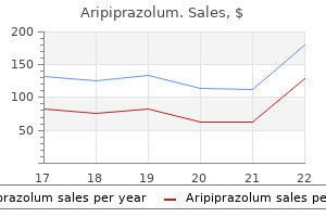
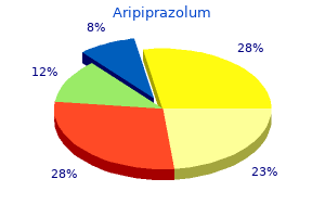
Aripiprazolum 10 mg discount free shipping
Tobacco and alcohol use depression definition and symptoms 10 mg aripiprazolum discount with amex, food plan depression hurts 10 mg aripiprazolum buy mastercard, and train symbolize the vast majority of things that influence preventable deaths. While behavioral adjustments are frequently troublesome to obtain, it must be emphasized that studies present even transient (<5 min) tobacco counseling by physicians leads to a major fee of long-term smoking cessation. The top causes of age-specific mortality and corresponding preventative strategies are listed in Table 205-1. In addition to the final recommendations relevant to all persons, screening for particular ailments and preventive measures need to be individualized based on family history, journey historical past, or occupational history. Specific suggestions for disease prevention can be present in subsequent chapters on "Cardiovascular Disease Prevention" (Chap. This determine and the footnotes describe indications for which vaccines, if not previously administered, must be administered unless famous in any other case. Many also confer herd immunity (an indirect effect), by which transmission of infections to unimmunized populations is lowered. Atherosclerotic cardiovascular disease is the main reason for death worldwide; prevention is targeted at modifiable risk factors (Table 207-1). Observational research present that smoking cessation reduces the chance of coronary occasions inside months. For pts who smoke, implement counseling and, as wanted, pharmacologic remedy to help cessation. Regular physical exercise, dietary improvements, and attaining fascinating body weight are beneficial for all pts with dyslipidemia. Treatment ought to be most aggressive in pts with established illness and in these on the highest threat as proven in Table 207-2, the cornerstone of which is statin therapy. Treatment of elevated blood pressure reduces the rate of stroke, congestive heart failure, and coronary occasions. Cardiovascular event charges in elderly pts with isolated systolic hypertension are additionally decreased by antihypertensive therapy. Dietary counseling, weight reduction, and increased physical activity are necessary in reducing the prevalence of this syndrome. Male Sex/Postmenopausal State Coronary risk is bigger in men in comparison with that of premenopausal girls of same age, however feminine danger accelerates after menopause. The antithrombotic drug aspirin reduces future extra cardiovascular occasions and mortality. Recent potential randomized trials have concluded that the cardiovascular advantages and bleeding danger of aspirin in main prevention closely match each other. Whether or not to prescribe aspirin for primary prevention should be based on a discussion with the pt about the potential advantages and risks in his/her specific case in order that a mutual decision can be made. Lifestyle Modifications Encourage beneficial train habits (>30 min average intensity bodily exercise daily) and sensible diet (low in saturated and trans fats; inclusion of fish, vegetables, complete grains, fruits; balance caloric consumption with vitality expenditure). Every physician go to is an opportunity to teach and reinforce the weather of a wholesome lifestyle. Cancer screening within the asymptomatic inhabitants at average risk is a complex concern. Screening may cause harm; problems may ensue from the screening test or the checks carried out to validate a optimistic screening take a look at or from remedies for the underlying illness. Evaluation of screening tools could be biased and must depend on potential randomized studies. Lead-time bias occurs when the pure historical past of disease is unaffected by the analysis, but the pt is recognized earlier in the midst of illness than regular; thus, the pt spends more of his/her life span figuring out the diagnosis. Length bias happens when slow-growing cancers which may never have come to medical consideration are detected during screening. Selection bias is the term for the truth that individuals who volunteer for screening trials may be totally different from the final inhabitants. Volunteers might need household history concerns that really elevate their threat, or they could be generally extra health-conscious, which can have an effect on consequence. The varied groups that evaluate and recommend screening apply tips have used varying standards to make their recommendations (Table 208-2). The absence of knowledge on survival for a quantity of ailments has led to an absence of consensus. Breast most cancers: the info on annual mammography help its use in girls age >50 years. Women age 40�49 years have a a lot lower incidence of breast cancer and a better falsepositive price on mammography. Nearly half of women screened throughout their forties will have a false-positive take a look at. Colon most cancers: Annual fecal occult blood testing after age 50 years is felt to be helpful. Commercial checks that combine detection of occult blood and the presence of genetic abnormalities in stool. However, 96% of the optimistic checks are false-positives and general survival is improved by only 6. Aromatase inhibitors have usually been superior to tamoxifen within the adjuvant remedy of hormone-sensitive breast most cancers, and certainly one of them (exemestane) reduces the chance of breast cancer by 65% in postmenopausal women at increased threat. Mutations in these genes carry a lifetime likelihood of >80% for creating breast most cancers. Bilateral prophylactic mastectomy prevents no less than 90% of these cancers however is a extra radical prevention than the usual treatment for the illness. Risk components embody diets excessive in saturated fats and low in vegetables and fruits, smoking, and alcohol consumption. Celecoxib, sulindac, and even aspirin seem to be efficient, and celecoxib is permitted by the U. The disease is very prevalent, with post-mortem studies finding prostate most cancers in 70�80% of males age >70. Surgical elimination, cryotherapy, or laser therapy is used to deal with the illness and is efficient in 80%. The vaccine is beneficial for all females and males age 9�26 years and will forestall as a lot as 70% of all cervical cancer. Initial research claimed that retinoids prevented the development of second primary tumors, a common function of head and neck cancer. Provide a transparent, sturdy, and customized message that smoking is a vital well being concern. A quit date should be negotiated within a number of weeks of the go to, and a follow-up contact by workplace workers around the time of the give up date should be supplied. Incorporation of cessation help into a practice requires a change of the care delivery infrastructure. These misconceptions perpetuate inadequate attention to modifiable threat components in girls, corresponding to dyslipidemia, hypertension, and cigarette smoking. Furthermore, since girls within the United States stay on common about 5 years longer than males, the majority of the disease burden for so much of age-related disorders.
10 mg aripiprazolum purchase with mastercard
Sinusoid feeding into the central venule Hepatic sinusoid Central venule Hepatocyte plates are formed by single rows of hepatocytes depression definition of 15 mg aripiprazolum buy mastercard. Lymphatic vessels encompass the blood vessels and bile ductules in the portal area anxiety 4th breeders buy 15 mg aripiprazolum. Kupffer cell Perisinusoidal cell Hepatocyte plate 2 Bile canaliculus 3 Canal of Hering Space of Mall Lymphatic vessel Portal venule Hepatic arteriole Bile duct Portal space Limiting plate 3 the canal of Hering (or cholangiole) is the terminal point of the community of bile canalicular trenches found on the hepatocyte surfaces. The canal of Hering is situated at the periphery of the hepatic lobule (periportal site), is lined by a squamous-to-cuboidal simple epithelium and connects with the bile ductules in the portal area after perforating the limiting plate. The connective tissue of the portal house supplies support to the portal triad, fashioned by branches of the hepatic artery (arteriole), portal vein (venule) and bile duct (ductule). In addition, lymphatic vessels and nerve fibers are current in the portal space (also designated portal canal, portal area or portal tract). Bile flows from the bile canaliculi into periportal bile ductules (cholangioles, or canals of Hering) after which into the bile ducts (or ductules) of the portal house after crossing the hepatic plate at the periphery of the hepatic lobule. Concepts of the hepatic lobule (17-12) There are three conceptual interpretations of the structure of the liver lobule: 1. The portal lobule idea, based mostly on the bile drainage pathway from adjacent lobules towards the same bile duct. The liver acinus idea, based mostly on the gradient distribution of oxygen along the venous sinusoids of adjoining lobules. The classic hepatic lobule is typically described as a polyhedral construction, often depicted as a hexagon with a central venule to which blood sinusoids converge. Components of the portal triad, branches of the portal vein and hepatic artery and a bile duct, are often found at the angles of the hexagon. However, recognition of the elements of the portal triad is helpful in determining the boundaries of the human hepatic lobule. In the portal lobule, the portal triad is the central axis, draining bile from the encompassing hepatic parenchyma. Functional concerns have modified the basic view and a liver acinus idea has gained floor in pathophysiology. In the liver acinus, the boundaries are decided by a terminal department of the hepatic artery. Glycogen Lipid droplet the smooth endoplasmic reticulum in hepatocytes is very developed and is at all times associated with clusters of glycogen molecules forming typical rosette-like inclusions. Stored glycogen in hepatocytes represents a glucose reserve for the maintenance of sugar concentrations in blood. Smooth endoplasmic reticulum Bile canaliculus Rough endoplasmic reticulum Nucleus Liver tissue stained with periodic acid�Schiff reagent to reveal deposits of glycogen (magenta staining) within the cytoplasm of hepatocytes. Rough endoplasmic reticulum Albumin, a major product of the hepatocyte, maintains plasma oncotic stress. Complement proteins, synthesized by hepatocytes, take part within the destruction of pathogens. Smooth endoplasmic reticulum the smooth endoplasmic reticulum has an important perform in detoxification. Enzymes essential for the detoxification of medicine (barbiturates), steroids, alcohol and different toxicants reside in the membrane of the smooth endoplasmic reticulum. Hepatocyte (17-13 to 17-17; see 17-11) Glycogen Zone I is the richest in oxygen and vitamins. Although pathology changes in the liver are usually described in relation to the traditional lobule, the liver acinus idea permits an understanding of liver regeneration patterns, liver metabolic actions and the event of cirrhosis. The perisinusoidal house of Disse separates the hepatocyte plates from the blood sinusoidal area. Kupffer cell is a differentiated phagocytic cell derived from monocytes A hepatocyte has distinct domains: apical domains, represented by the bile poles, and in depth basolateral domains with microvilli extending into the space of Disse. Basolateral domain Apical domain Golgi apparatus Fenestrated endothelium Reticular fibers Space of Disse Nucleus Bile canaliculus Apical domain Peroxisome Rough endoplasmic reticulum Peroxisome A membrane-bound structure that contains oxidases and catalase. Basolateral area Lipid droplet Nucleus Smooth endoplasmic reticulum and related glycogen inclusions Bile canaliculus the bile canaliculus is an extracellular canal between adjacent hepatocytes. Bile launched into the canaliculus is drained by the canal of Hering, or cholangiole, an epithelial-lined ductule in the periportal area. The canal of Hering carries the bile to the bile ductules, one of the three elements of the portal house. Endothelial cells have a fenestrated cytoplasm associated with a discontinuous basal lamina. The space of Disse, between the sinusoid and the basolateral domain of hepatocytes, permits an change between blood and hepatocytes. Hepatocyte absorptive operate is enhanced by the microvilli extending into the space of Disse. Fenestrated endothelial cell lining of a hepatic sinusoid Microvilli of the basolateral area of a hepatocyte, extending into the subendothelial area of Disse Rough endoplasmic reticulum Glycogen the bile canaliculus is an area restricted by two or more hepatocytes. Tight junctions seal the intercellular house, thus stopping the leakage of bile. Microvilli Lumen of the bile canaliculus Occluding junction in connective tissue, are separated from the hepatic lobule by a limiting plate of hepatocytes (see 1711). Blood from the portal vein and hepatic artery flows into the sinusoids and is drained by the central venule. Lymph flows from the area of Disse to a lymphatic vessel also within the portal area (see 17-13). Gap junctions on the lateral surfaces of adjoining hepatocytes enable intercellular useful coupling. Excess fluid within the area of Disse is collected within the house of Mall, located on the periphery of the hepatic lobule. Lymphatic vessels piercing the limiting plate drain the fluid of the space of Mall. Note that hepatocytes synthesize several plasma proteins required for blood clotting (see Chapter 6, Blood and Hematopoiesis). The apical area borders the bile canaliculus, a trench-like melancholy lined by microvilli and sealed on the sides by occluding junctions to prevent leakage of bile, the exocrine product of the hepatocyte. The hepatocyte contains tough endoplasmic reticulum, involved within the synthesis of plasma proteins and a extremely developed easy endoplasmic reticulum, associated with the synthesis of glycogen, lipid and detoxification mechanisms. Enzymes inserted in the membrane of the smooth endoplasmic reticulum are involved within the following capabilities: 1. The removal of iodine from the thyroid hormones triiodothyronine (T3) and thyroxine (T4). The detoxing of lipid-soluble medicine similar to phenobarbital, throughout which the graceful endoplasmic reticulum is considerably developed. The Golgi equipment contributes to glycosylation of secretory proteins and the sorting of lysosomal enzymes. Lysosomes degrade aged plasma glycoproteins internalized at the basolateral area by a hepatic lectin membrane receptor, the asialoglycoprotein receptor, with binding affinity to terminal galactose after the removing of sialic acid.
Safe 20 mg aripiprazolum
It is involved within the synthesis of proteins mood disorder quizzes 20 mg aripiprazolum buy otc, carried out by their hooked up ribosomes depression test learnmyself order aripiprazolum 20 mg on line. Most proteins exit the rough endoplasmic reticulum in vesicles transported to the cis portion of the Golgi equipment. Leaflets of cytomembranes and plasma membrane the luminal compartment of a secretory cell is steady with the exterior of the cell the exocytoplasmic leaflet faces the luminal compartment Protoplasmic leaflet Exocytoplasmic leaflet Secretory granule 2 Transporting vesicle 1 Endoplasmic 3 Golgi reticulum compartment equipment 4 Secretory vesicles 5 Exocytosis Plasma membrane Endoplasmic reticulum Cytosol Golgi apparatus Cytosol the protoplasmic leaflet faces the cytosolic compartment Freeze-fracture method (Primer 2-A) the freeze-fracture approach is efficacious for the visualization of intramembranous proteins with the electron microscope. This method offered the first evidence for the presence of transmembrane proteins within the plasma membrane and cytomembranes. Specimens are frozen at liquid nitrogen temperature (�196oC) and "break up" with a knife (under high vacuum) along the hydrophobic core of the membrane. As a result, two complementary halves, corresponding to every membrane bilayer, are produced. A replica of the specimen is generated by evaporating a very thin layer of a heavy steel (generally platinum with a thickness of 1. The platinum replica is then detached from the true specimen by floating it on a water floor, mounted on a steel grid and examined underneath the electron microscope. Primer 2-A signifies the nomenclature for the identification of surfaces and faces in electron micrographs of freeze-fracture preparations. One last point: Note that a transmembrane protein stays with the protoplasmic leaflet, leaving a complementary pit in the opposite exocytoplasmic leaflet. Cortical cytoskeletal parts are directly or indirectly hooked up to the tip of the protein uncovered to the cytoplasmic side of the plasma membrane and play an active half within the upkeep and transforming of protein and lipid domains. When cells had been labeled with a radioactive amino acid to hint the intracellular pathway of the secreted proteins, it was found by autoradiography that, after a 3-minute labeling, newly synthesized proteins had been localized in the rough endoplasmic reticulum 1. Later on, the radiolabeled proteins were found to translocate to the Golgi apparatus 2 and then, inside secretory vesicles as zymogen granules 3, to the plasma membrane and the extracellular house 4. Golgi equipment (2-12 and 2-13) the Golgi equipment consists of a cluster of flattened stacks of sacs called cisternae stabilized by golgins, a household of coiled-coil proteins. Cisternae of the medial-Golgi are interposed between the cis-Golgi and the trans-Golgi. Cargos derived from the endoplasmic reticulum transport soluble proteins and membrane to the cis-Golgi. Cargo designates newly synthesized membrane and proteins destined to be stored inside a cell compartment or secreted to the cell exterior. The material travels through the cisternae by means of transport vesicles that bud off from one cisterna and tether and fuse with the subsequent in the presence of golgins. Golgins type an appendicular community on the cis-Golgi, around the rims of the sacs and on the trans-Golgi, with roles in Golgi construction stabilization and vesicle trafficking (see 2-13). Functions of the Golgi equipment (see 2-13) Three specific features are carried out by the Golgi equipment: 1. Modification of carbohydrates hooked up to glycoproteins and proteoglycans obtained from the endoplasmic reticulum. A characteristic glycosylation event throughout the Golgi is the modification of N-linked oligosaccharides on glycoproteins. After protein synthesis, transmembrane proteins stay anchored to the membrane of the endoplasmic reticulum cisterna by one or more hydrophobic transmembrane segments as a consequence of stop-transfer signals (not shown). These signals stop the whole translocation of a protein throughout the membrane. The enzymes referred to as glycosyltransferases add particular sugar residues; the enzymes called glycosidases remove particular sugar residues. We talk about in another section of this chapter how the Golgi equipment marks specific proteins for sorting to lysosomes. Once processed, cargos bud off from the Golgi apparatus and are either sorted to the secretory of lysosomal sorting pathway (anterograde traffic) or back to the endoplasmic reticulum (retrograde traffic). Certain classes of cargos are saved into secretory 80 granules for later release in response to an extracellular sign. This mechanism is called constitutive secretion; it supplies newly synthesized lipids and proteins to the plasma membrane or proteins to be released exterior the cell, such as proteins of the extracellular matrix or immunoglobulins throughout immune reactions. Cargo sorting happens along microtubules or actin filaments with the help of motor proteins. The presence of particular lipids domains in the membrane of a vesicle cargo recruit coating proteins and tethering golgins to type the cargo within the path of an acceptor membrane website. The saccules and vesicles contain proteins being glycosylated for additional secretion or sorting. The Golgi equipment consists of 4 major functionally distinct compartments: (1) the cis-Golgi is the entry website to the Golgi apparatus of products derived from the endoplasmic reticulum. When the vesicle cargo reaches an acceptor membrane, it fuses with the assistance of fusion proteins. Vesicle transport (2-13) Vesicle transport involves the mobilization of proteins and membrane between cytomembrane compartments. The exocytosis or secretory pathway starts within the endoplasmic reticulum, continues through the Golgi apparatus and ends on the cell floor. The endocytic pathway consists of the internalization and degradation of extracellular materials from the plasma membrane, by way of endosomes to lysosomes. These two events depend on distinctive proteins coating the cytosolic side of the membrane of the transport vesicle, which turns into a coated vesicle. Clathrin-coated vesicles are seen in the exocytosis/ secretory and in the endocytosis pathway. In the endocytosis pathway, vesicles begin on the plasma membrane as clathrin-coated pits. Clathrin molecules assemble in a basket-like association on the cytosolic face of the plasma membrane and the pit shape modifications right into a vesicle. Adaptins stabilize the clathrin coat to the vesicle membrane and assist within the choice of cargos for transport by binding to cargo receptors on the vesicle membrane. When the cargo reaches the goal acceptor membrane, the coat proteins are shed and the membranes can fuse. Lysosomal sorting pathway (2-14) the cisterna closest to the endoplasmic reticulum is the cis-Golgi, whereas the cisterna closest to the apical domain of the cell is the trans-Golgi. Transporting vesicles bud from one stack and fuse with the subsequent in an anterograde traffic (endoplasmic reticulum to Golgi) or retrograde traffic (Golgi to endoplasmic reticulum). The insertion in the cis-Golgi of mannose6-phosphate (M6P) into oligosaccharides attached to glycoproteins focused to lysosomes. By this mechanism, M6P-containing lysosomal enzymes are separated from different glycoproteins in vesicles with the M6P receptor. After being transported to a clathrin-coated transporting vesicle, lysosomal enzymes dissociate from the M6P receptor and turn out to be surrounded by a membrane to form a lysosome. Membranes containing free M6P receptor are returned to the Golgi equipment for recycling.
Buy aripiprazolum 20 mg with amex
The main goals are to erase and reprogram methylation patterns to reset imprints and/or eliminate acquired epigenetic modifications anxiety ed aripiprazolum 20 mg purchase otc. Both syndromes have an epigenetic defect caused by an absence of methylation of a quantity of paternally derived alleles anxiety treatment centers discount 10 mg aripiprazolum. After examining a number of serial cross sections covering a distance of a few millimeters or centimeters, we decide the successive mobile associations (or stages of a cycle) alongside the length of the seminiferous tubule. The distance separating two similar stages defines the length of a spermatogenic wave. The variety of levels that constitute a spermatogenic cycle and the variety of cycles required for the completion of a spermatogenic progeny vary among species. The variety of cellular associations or stages in a cycle is constant for any given species (14 levels within the rat, 6 phases in man, 12 in the monkey). In human testes, spermatogenic cell progenies are organized in a helical style, as a substitute of a longitudinal association as in rodents (see 20-16). Consequently, a cross section of a seminiferous tubule will show three to 4 cellular associations, instead of the single one observed within the testes of rodents. In our hypothetical instance proven in 20-17, each of the 4 cycles consists of 6 consecutive phases, which reappear repeatedly. Changes in chromatin structure from a nucleosomal-type to a smooth-type of chromatin in late spermatids (see 20-10). Paternal chromatin undergoes main remodeling because of the chromatin-associated protamine molecules (acquisition of histones). Inner cell mass accommodates pluripotent cells capable of give rise to all cell sorts within the embryo, in addition to the extra-embryonic cells. During gametogenesis (spermatogenesis and oogenesis), genetic imprints are differentially erased to permit epigenetic reprogramming to be transmitted by the gametes to embryos. In abstract, reprogramming throughout gametogenesis is required for the resetting of imprints or eliminating acquired epigenetic modifications. Epigenetic reworking happens shortly after fertilization and totipotency is restored to the zygote. Which are the underlying molecular mechanisms enabling primordial germ cells to turn into gonocytes resulting in gametogenesis During gametogenesis, the differential expression of alleles (Greek allos, another) could be inhibited in paternal and maternal gametes. As you remember from earlier discussions, genes are available pairs, one copy or allele inherited from every parent. During spermatogenesis and oogenesis one copy of the imprinted gene is selectively silenced. Imprinting problems are seen when the choice maternal or paternal copy (allele) is disrupted. Prader-Willi syndrome is characterized by hypotony, respiratory distress, obesity, quick stature and gentle psychological disability. It is attributable to the deletion of a paternal allele or the retention of two maternal copies. Histone deacetylase relocates to the histone core and removes acetyl groups from histones. Histone methyltransferase capability, extreme inappropriate laughter, lack of speech and hyperactivity. In distinction to Prader-Willi syndrome, the maternal allele has been misplaced or two paternal copies are retained. With this background, we now tackle the molecular features of epigenetics reprogramming (see 20-20). A large number of CpG islands are current in transcription initiation websites and in promoters of energetic genes. Chromatin of an actively transcribing gene (euchromatin) has acetylated histones and the CpG islands are unmethylated. Chromatin may be 694 condensed (heterochromatin) to turn out to be transcriptionally inactive. Histone deacetylases take away acetyl teams from the N-terminal tail of the nucleosomal histones. Methylation consists of the attachment of a methyl group to a organic molecule by methyltransferases. Histone deacetylation is a prerequisite for histone methylation, which includes histone methyltransferase targeting histone three (H3). The management of these sufferers is increasingly essential, particularly with regard to their sexuality and fertility. Malignant cells resembling seminoma cells are confined inside seminiferous tubules. Each testis is surrounded by the tunica albuginea (dense connective tissue) concentrated in the mediastinum, where the rete testis is positioned. The network of blood vessels beneath the tunica albuginea known as tunica vasculosa. Septa or partitions derived from the mediastinum divide the testes into 250 to 300 lobules. Teratoma is a benign germ cell tumor derived from a combination of the tissues from all three embryonic layers (ectoderm, mesoderm and endoderm). The tumor consists of cysts (containing mucoid material and nodules of cartilage), stable tissue (immature form) and malignant reworked teratomas. In distinction to germ cell tumors, it presents metastasis before a testicular mass is found. Yolk sac tumor is the commonest testicular tumor of childhood and younger patients. Typical seminoma could mimic a Sertoli cell tumor because of the microlobular group and presence of cells with clear nuclei and conspicuous nucleolus like Sertoli cells. A basement membrane, shaped by a basal lamina and reticular lamina, separates the wall from the seminiferous epithelium. The two ends of the seminiferous tubule open into the rete testis, a network of channels amassing testicular sperm, secretory proteins and fluid produced by the seminiferous epithelium. It accommodates blood vessels, lymphatic channels and clusters of the androgen-producing Leydig cells. The stratified mobile association of spermatogenic cells (spermatogonia, main and secondary spermatocytes and spermatids) lends to the classification of the seminiferous epithelium as stratified with structural and functional characteristics not found in another stratified epithelia. For instance, a everlasting postmitotic cell inhabitants of somatic Sertoli cells interacts with transient mitotically dividing spermatogonia, meiotically dividing spermatocytes and differentiating haploid spermatids. The everlasting member of the epithelium, the Sertoli cell, maintains a physical and useful relationship with all the transient members, the spermatogenic cells.
Aripiprazolum 20 mg free shipping
Numerous osteogenesis centers develop and finally fuse mood disorder screening tool cheap 10 mg aripiprazolum fast delivery, forming a community of anastomosing trabeculae resembling a sponge depression from work generic aripiprazolum 10 mg with visa, the so-called spongy bone or primary spongiosa. Because collagen fibers in the newly formed trabeculae are randomly oriented, the early intramembranous bone is described as woven bone, in contrast with the frequently oriented collagen fibers of the lamellar or compact bone shaped later throughout bone remodeling. Calcium phosphate is deposited within the bone matrix or osteoid, which turns into mineralized. Osteocytes stay related to one another by cytoplasmic processes enclosed within slim tunnels referred to as canaliculi. New osteoblasts are generated from preosteoblasts, the osteoprogenitor cells, positioned adjacent to the blood vessels. In lamellar bone, the newly synthesized collagen fibers are aligned into bundles with a regular orientation. Lamellae, organized in concentric rings round a central haversian canal occupied by a blood vessel, type osteons, or haversian techniques. Membranous bones stay as spongy bone within the center, the diplo�, enclosed by an outer and an inner layer of lamellar compact bone. The condensation of the external and internal connective tissue layers to kind the periosteum and endosteum, respectively, containing osteoprogenitor cells. Endochondral ossification (5-3 to 5-10) Endochondral ossification is the method by which skeletal cartilage templates are replaced by bone. Bones of the extremities, vertebral column and pelvis (the appendicular skeleton) derive from a hyaline cartilage template. As in intramembranous ossification, a major bone growth middle is formed during endochondral ossification (see 5-3). Shortly thereafter, chondrocytes in the central region of the cartilage bear hypertrophy and synthesize type X collagen, a marker for hypertrophic chondrocytes. Hypertrophic chondrocytes undergo apoptosis as calcification of the matrix in the midst of the shaft of the cartilage template takes place. At the identical time, the inside perichondrial cells exhibit their initial bone improvement potential and a skinny periosteal collar of bone is shaped across the midpoint of the shaft, the diaphysis. Consequently, the first bone development middle finally ends up situated inside a cylinder of bone. The periosteal collar, shaped under the periosteum by apposition, consists of woven bone. This course of is controlled by patterning signals from polypeptides of the Wnt, hedgehog, fibroblast growth factor and reworking progress factor� families. Osteocytes within the core of the blastema are interconnected by cell processes forming a practical syncytium. Later, Ca2+, transported by blood vessels, is used in the mineralization process and first bone tissue is shaped. Ca2+ 3 2 Osteocyte Blood vessel Mesenchymal cell Bone matrix (osteoid) Osteoblast Osteoclast Mineralization Organization of a major middle of ossification Multiple individual trabeculae enlarge by appositional development and finally fuse together as a major osteogenesis heart organized through the first stage of intramembranous ossification. Although major bone tissue formation begins as an Blood vessel interstitial course of, it quickly turns into appositional. At the surface of the osteoid, osteoblasts continue the appositional deposit of matrix, mainly kind I collagen and non-collagenous proteins. Blood vessels invade the area formerly occupied by the hypertrophic chondrocytes they usually branch and project toward either end of the middle of bone growth. Osteoprogenitor cells (preosteoblasts) and blood growth stem cells reach the core of the calcified cartilage by way of the perivascular connective tissue surrounding the invading blood vessels. Then, preosteoblasts differentiate into osteoblasts that aggregate on the surfaces of the calcified cartilage and start to deposit bone matrix (osteoid). At this developmental step, a major center of ossification, outlined by the periosteal collar and the center of ossification in the inside of the cartilage template, is organized on the diaphysis. Intramembranous ossification is characterized by: A well-vascularized primitive connective tissue. An combination of mesenchymal stem cells differentiates directly into osteoid-producing osteoblasts. The mesenchymal cells situated near the surface condense to kind the periosteum Blood vessel Monolayer of osteoblasts the continued deposition of bone on trabecular surfaces determines the occlusion of the intertrabecular areas, and compact bone is shaped. Osteoblasts manage thin trabeculae of woven bone, forming an irregular community referred to as primary spongiosa. Blood vessel Acidophilic osteoid Trabecula whereas the center of the cartilage is being replaced by bone on the equidistant zones of ossification. After birth, secondary facilities of ossification develop within the epiphyses (see 5-4). As within the diaphysis, a hundred and eighty the house occupied by hypertrophic chondrocytes is invaded by blood vessels and preosteoblasts from the perichondrium. Most of the hyaline cartilage of the epiphyses is changed by the spongy bone, aside from the articular cartilage and a skinny disk, the epiphyseal growth plate, located between the epiphyses and the diaphysis. The epiphyseal progress plate is answerable for subsequent progress in length of the bone by a mechanism that we talk about later. Hypertrophic chondrocytes secrete vascular endothelial cell growth factor to induce sprouting of blood vessels from the perichondrium. Then, calcification of the matrix and apoptosis of hypertrophic chondrocytes happen. Blood vessels, forming the periosteal bud, department in opposite directions Zones of endochondral ossification (see 5-3 to 5-8) We have seen that the deposition of bone within the center of the diaphysis is preceded by an erosion process in the hyaline cartilage template (see 5-3 and 5-4). This middle of abrasion, defined as the first ossification center, extends in opposite directions of the template, coinciding with the formation of a bony collar. The bony collar supplies power to the midsection of the diaphysis or shaft because the cartilage is weakened by the gradual elimination of the cartilage before its substitute by bone. The continuing means of cartilage erosion and bone deposition may be visualized histologically. Four main zones can be distinguished in 5-5, starting on the finish of the cartilage and approaching the zone of erosion: 1. The reserve zone is a web site composed of primitive hyaline cartilage and is answerable for the expansion in size of the bone as the erosion and bone deposition process advances. The proliferative zone is characterized by the energetic mitotic exercise of chondrocytes aligning as mobile stacks parallel to the long axis of the cartilage template (see 5-6 and 5-7). The hypertrophic zone is outlined by chondrocyte apoptosis and calcification of the territorial matrix surrounding the columns of beforehand proliferated chondrocytes. Despite their structurally collapsing look, postmitotic hypertrophic chondrocytes play an necessary role in bone development. To instruct adjacent chondrocytes of the perichondrium to change into preosteoblasts and continue forming the bone collar.
Purchase 10 mg aripiprazolum with visa
The phase past the arteriole proper is the metarteriole depression symptoms on the body aripiprazolum 10 mg discount amex, the terminal department of the arterial system mood disorder with anxiety icd 9 order aripiprazolum 10 mg mastercard. It consists of one layer of muscle cells, typically discontinuous, and represents an necessary local regulator of blood flow. Capillaries (exchange vessels) (12-7) Arterioles, capillaries and venules represent the microvasculature, the positioning where most intercellular communication happens. Arterioles regulate the distribution of blood to totally different capillary beds by vasoconstriction and vasodilation in localized regions. The regulation of the vascular tone (partial contraction of the vascular easy muscle) is an arterial function that happens primarily at the level of the arterioles. Arterioles are structurally tailored for vasoconstriction and vasodilation because their partitions comprise circularly arranged clean muscle fibers. Arterioles are regarded as resistance vessels and are the most important determinants of systemic blood strain. The diameter of arterioles and small arteries Blood capillaries are extremely skinny tubes shaped by a single layer of highly permeable endothelial cells surrounded by a basal lamina encased by pericytes. Pericytes make contacts with the basal lamina and sometimes with endothelial cells, to regulate vessel stability and transendothelial transport. However, collecting lymphatic vessels have a easy muscle cell layer that allows pumping by undergoing phasic and tonic contractions. The diameter vary of a blood capillary is about 5 to 10 m, massive sufficient to accommodate one purple blood cell and skinny sufficient (0. External elastic lamina Lumen Blood vessels Lymphatic Nerve Tunica adventitia Loose connective tissue, blood vessels (vasa vasorum), lymphatics and nerves (nervi vasorum) Lumen Red blood cell (lumen) Endothelium Internal elastic lamina Functional characteristics of muscular arteries Arteries conduct blood from the center to the capillaries and likewise retailer a portion of the ejected blood during every cardiac systole to enable the flow to proceed through the capillaries during cardiac diastole. When blood stress is decided in a person by a sphygmomanometer, systolic stress is recorded by a stethoscope as a tapping sound originating within the artery distal to the cuff. When the cuff stress decreases beneath the height arterial strain (below a hundred and twenty mm Hg), spurts of blood move via the externally compressed artery. Diastolic pressure is recorded when the tapping sound disappears as the cuff strain falls beneath minimal arterial strain (below eighty mm Hg). Tunica media Smooth muscle cell Elastic lamella Tunica adventitia Vasa vasorum culation, is composed of the terminal arteriole (and metarteriole), the capillary bed and the postcapillary venules. The capillary mattress consists of slightly giant capillaries (called preferential or thoroughfare channels), the place blood flow is continuous, and small capillaries, called the true capillaries, the place blood circulate is intermittent. The amount of blood entering the microvascular mattress is regulated by the contraction of smooth muscle cells of the precapillary sphincters. The sphincters are situated where true capillaries arise from the arteriole or metarteriole. When functional demands decrease, most precapillary sphincters are closed, forcing the circulate of blood into thoroughfare channels. Postcapillary venules are the first sites of 426 leukocyte extravasation throughout inflammation. Arteriovenous shunts, or anastomoses, bypass the microvascular bed by connecting instantly arterioles to postcapillary venules. The native conditions of the tissues (concentration of nutrients and metabolites and different substances) can control native blood move in small portions of a tissue area. Types of capillaries (12-8 and 12-9) Three morphologic forms of capillaries are acknowledged (see 12-8): 1. The cytoplasm of vascular smooth muscle cells accommodates actin and myosin filaments whose contraction is managed by calcium. Arteriolar clean muscle cells contract in response to increased transmural strain and relax when the stress decreases. Pericytes are undifferentiated cells that resemble modified smooth muscle cells and are distributed at random intervals in close contact with the basal lamina. Endothelial cells are linked by tight junctions and transport fluids and solutes by caveolae and pinocytotic vesicles. Fenestrated capillaries have pores, or fenestrae, with or with out diaphragms (see 12-9). Fenestrated capillaries with a diaphragm are found in intestines, endocrine glands and round kidney tubules. Fenestrated capillaries and not utilizing a diaphragm are characteristic of the renal glomerulus. In this specific case, the basal lamina constitutes an necessary permeability barrier. Discontinuous capillaries are characterized by an incomplete endothelial lining and basal lamina, with gaps or holes between and within endothelial cells. Discontinuous capillaries and sinusoids are found where an intimate relation is required between blood and parenchyma (for instance, in the liver, spleen and bone marrow). Arterial and venous portal techniques (12-10) In general, blood from an arteriole flows right into a capillary community and is drained by a venule. There are two specialized capillary methods that depart from this commonplace arrangement: 1. In the kidneys, an afferent arteriole drains into a capillary community called the glomerulus. The glomerular capillaries coalesce to type an efferent arteriole, which branches into another capillary network known as the vasa recta. The vasa recta encompass the limbs of the loop of Henle and play a significant role in the formation of urine. In the portal system, intestinal capillaries are drained by the portal vein to the liver. In the liver, the portal vein branches into venous sinusoids between cords of hepatocytes. Blood flows from the sinusoids right into a collecting vein after which back to the heart by way of the inferior vena cava. Venules join the primary sinusoidal plexus of the hypothalamus (median eminence) with the secondary plexus in the anterior lobe of the hypophysis, forming the hypophyseal-portal system. This system transports releasing components from the hypothalamus to stimulate the secretion of hormones into the bloodstream by cells of the anterior hypophysis. Open or closed precapillary sphincters can regulate the blood circulate throughout the capillary bed. Sympathetic nerve Terminal arteriole (derived from a small arteriole) Smooth muscle cell Venule Blood move via true capillaries is intermittent and managed by the constriction of the arteriole or precapillary sphincters. A preferential or thoroughfare channel allows steady blood move from the arteriole to the postcapillary venule. True capillaries Pericyte A metarteriole can function a thoroughfare channel to the postcapillary venule (bypassing the capillary bed), or as a conduit to supply the capillary bed. Postcapillary venule (without smooth muscle cells) Venule (with wrapped smooth muscle cells) Venule Smooth muscle cells Arteriole Capillary Arterioles have an endothelial lining, a thick smooth muscle layer, and a thin adventitial layer. Arterioles can give rise to capillaries or, in some tissues, to metarterioles, which then give rise to capillaries. Arterioles regulate blood move via capillaries by constriction or dilation of the precapillary sphincters. Capillaries are quite a few in metabolically active tissues (such as cardiac and skeletal muscle and glands).
Aripiprazolum 15 mg without prescription
The lamina propria is composed of smooth muscle and elastic and collagenous fibers depression symptoms sleep 15 mg aripiprazolum buy with amex. Alveolar outpocket four Alveoli four Respiratory bronchioles the presence of alveolar outpocketings interrupt the continuity of the wall of the bronchiole clinical depression symptoms yahoo answers buy cheap aripiprazolum 20 mg online. The low cuboidal epithelium is replaced discontinuously by squamous sort I alveolar epithelial cells. The pores are liable for collateral respiration when blockage of a small bronchiole occurs. Thus, in case of a blockage, adjoining unobstructed bronchioles and associated alveoli continue to provide alveolar ventilation via the pores of Kohn. In this preparation stained with orcein, darkish blue�stained elastic fibers are seen along the wall of the terminal bronchiole and the respiratory bronchiole as properly as within the wall of a department of the pulmonary artery (tunica interna and adventitia). The lining epithelium of the terminal bronchiole accommodates membership cells (Clara cells) troublesome to resolve at low magnification. The intrapulmonary segmentation leads to the group of a pulmonary lobule and a pulmonary acinus. Pulmonary lobule and pulmonary acinus (13-9 and 13-10) A terminal bronchiole and the associated region of pulmonary tissues that it supplies constitute a pulmonary lobule (see 13-9). A pulmonary lobule includes the respiratory bronchioles, alveolar ducts, alveolar sacs and alveoli. The pulmonary acinus, the unit of gasoline change of the lung, is equipped by a respiratory bronchiole. Emphysema is permanent enlargement of the air areas distal to the terminal bronchioles, associated with the destruction of their partitions. The respiratory bronchiole and the initial portion of the alveolar duct show an interrupted wall with typical easy muscle knobs and scattered elastic fibers bulging into the lumen (see 13-9 and 13-10). At the distal finish of the alveolar duct, the sleek muscle knobs disappear and the liner epithelium is primarily kind I alveolar epithelial cells. At the alveolar duct, muscle knobs are coated by alveolar capillaries, which, in turn, are lined initially by type I alveolar cells. Following airway injury, membership cells proliferate and migrate to replenish alveolar epithelial cells. In addition, proliferative membership cells can produce ciliated cells and extra membership cells. Respiratory portion of the lung (13-12) Terminal bronchioles give rise to three generations of respiratory bronchioles (0. Respiratory bronchioles are the transition from the conducting to the respiratory portion of the lung. The alveolus (13-13 to 13-15; see 13-10 and 13-12) About 300 million air sacs, or alveoli, in each lung provide a total floor space of 75 m2 for oxygen and carbon dioxide exchange. The alveolar epithelium consists of two distinct cell sorts (see 13-12 and 13-13): 1. They derive from bone-marrow monocytes and are incessantly seen within the alveolar lumen and interstitium. Alveolar dendritic cells (see 13-15), actively monitor the alveolar air house for antigens and take them up for presentation to T cells. After airway harm, club cells can proliferate to regenerate the bronchiolar epithelium and even migrate to replenish alveolar epithelial cells. At the electron microscope level, the apical dome-shaped area of membership cells contains cytoplasmic dense secretory granules, mitochondria and numerous vesicles. Alveolus Alveolus uct ar d eol Alv Ciliated cell Alveolus Obliterative bronchiolitis or constrictive bronchiolitis Peribronchiolar inflammatory cell infiltrate the histologic sample of obliterative bronchiolitis of a terminal bronchiole is suitable with abnormal tissue repair and irritation in response to tissue injury, including poor epithelial regeneration, peribronchiolar fibroproliferation and continual Reduction of the inflammatory cell infiltrate. Obliterative bronchiolitis is a frequent complication Peribronchiolar fibrosis of hematopoietic stem-cell transplantation or lung transplantation. Remnants of the muscle knobs lined by a low cuboidal-to-squamous easy epithelium can be seen at the alveolar openings. Air circulate 3 Saccular outpocketings 2 Muscle knob: Smooth muscle cell bundles (muscle knobs), innervated by parasympathetic nerve fibers, contract to constrict the lumen of the bronchiole. In bronchial asthma, muscle contraction, triggered by histamine release from mast cells, is persistent. In addition to capillaries, the interstitium incorporates elastic and collagen fibers produced by interstitial fibroblasts. Alveolar cells are separated from capillary endothelial cells by the associated basal laminae produced by them. Multiple small perivascular lymphatics are liable for maintaining fluid balance in the alveolar interstitium. The cytoplasm shows dense membrane-bound lamellar our bodies, representing secretory granules containing pulmonary surfactant. Surfactant is released by exocytosis and spreads over a skinny layer of fluid that usually coats 464 the alveolar surface (see 13-16). By this mechanism, the pulmonary surfactant lowers the floor pressure at the air-fluid interface and thus reduces the tendency of the alveolus to collapse on the end of expiration. As previously indicated, club cells, located in terminal bronchioles, additionally secrete pulmonary surfactant. Alveolar macrophages are important for catabolizing the surfactant that lines the floor of the alveoli. There are two totally different sources of alveolar macrophages: (1) Self-renewal macrophages derived from a fetal yolk sac progenitor. Alveolar capillaries form an interconnected network round alveoli and are lined by steady endothelial cells, related to one another by occluding junctions. The endothelial basal lamina fuses with the basal lamina of the alveolar epithelium, where the alveolar capillary is carefully related to the alveolar wall (the favorable web site for gas exchange). At this site, fluids can move from capillaries to the interstitium, to drain eventually into para-alveolar lymphatics adjacent to an arteriolar wall comparable to branches of the pulmonary or bronchial arteries. In patients with heart illness, alveolar macrophages contain many vacuoles full of hemosiderin, resulting from phagocytosis of red blood cells and degradation of their hemoglobin. Alveolar macrophages are sentinel cells migrating over the luminal floor of the alveolus. These cells monitor any inhaled dust or micro organism that may have escaped entrapment by the mucous lining in the airway. When stimulated by metabolic products of micro organism, macrophages release chemotactic elements that induce transendothelial migration of leukocytes, which be a part of macrophages to neutralize invading microorganisms. An excess of asbestos (composed of silica, iron, sodium, magnesium and different metals) causes macrophages to launch chemical agents, producing alveolitis and eventual fibrosis of Alveolar the lung. Surfactant turnover is facilitated by the phagocytic perform of alveolar macrophages. Macrophages can take up inhaled asbestos and set off asbestosis (interstitial pulmonary fibrosis; see 13-17), the deposition of collagen and asbestos our bodies. The subsequent step is to explore how normal structure and performance of the airways are disrupted during illness. Asthma, to emphasize how mucus hypersecretion and bronchoconstriction interface with the immune system. Chronic bronchitis and emphysema, to strengthen the idea of pulmonary lobule and pulmonary acinus by exploring two examples of chronic obstructive pulmonary disease.
Cheap aripiprazolum 20 mg free shipping
Cancer stem cells specific stemness-associated genes depression criteria 20 mg aripiprazolum buy visa, such as Nanog mood disorder social security disability cheap aripiprazolum 20 mg on-line, Oct4, Myc, Sox2 and Klf4 (Kr�pel-like factor 4). Remember that stemness is the characteristic gene expression sample of various stem cells not noticed in strange, non-stem cells. The deposit of collagen and extracellular matrix components increases, resulting in a progressive fibrosis of the liver, a typical function of cirrhosis. An increased deposit of collagen fibers and extracellular matrix inside the house of Disse is followed by a loss of fenestrations and gaps of sinusoidal endothelial cells. As the fibrotic course of advances, myofibroblasts constrict the lumen of the sinusoids and improve vascular resistance. An improve in resistance to the move of portal venous blood within the hepatic sinusoids results in portal hypertension in cirrhosis. In summary, hepatocytes have regenerative functionality in response to harm and macrophages secrete matrix metalloproteases that break down scar tissue and enhance hepatocyte proliferation. However, the liver extracellular matrix controls the epithelial regenerative responses. Fibrogenesis disrupts the regenerative potential of hepatocytes and biliary epithelial cells to the purpose that liver regeneration is compromised. The vascular constructions become abnormal, collagen bundles encompass hepatocytes and cirrhosis develops. Long-term consumption of ethanol results in fatty liver (a reversible course of if ethanol consumption is discontinued), steatohepatitis (fatty liver accompanied by an inflammatory reaction), cirrhosis (collagen proliferation or fibrosis) and hepatocellular carcinoma (malignant transformation of hepatocytes). Injury of hepatocytes results in programmed cell death, or apoptosis, attributable to the activation of caspases (see Chapter 3, Cell Signaling Cell Biology Pathology). Alcohol is oxidized to acetaldehyde within the cytoplasm and acetaldehyde is transformed to acetate in mitochondria. An extra of H+ and acetaldehyde causes mitochondrial injury, disrupts microtubules and alters proteins that may induce autoimmune responses, resulting in hepatocyte injury. Reactive oxygen produces harm to hepatocytes by causing lipid peroxidation, resulting in cell membrane injury. Hepatocyte harm Smooth endoplasmic reticulum Excess of oxygen radicals Ethanol Large fats deposits within the cytoplasm of hepatocytes are observed in fatty liver (steatosis) following long-term consumption of alcohol. Patients with chronic forms of viral hepatitis, lasting greater than 6 months, can transmit the infection to others with blood or physique fluids, and the infection can evolve over time to cirrhosis or result in the development of hepatocellular most cancers (liver cancer). The advanced is water-soluble and can enter the brain to trigger extreme neurologic issues (kernicterus) in hemolytic disease of the new child (erythroblastosis fetalis). Unconjugated bilirubin is launched from the macrophage and reaches the blood circulation. Excessive production of unconjugated bilirubin, resulting from excessive destruction of red blood cells, results in jaundice. The bilirubin-ligandin advanced reaches the smooth endoplasmic reticulum and free bilirubin is released into the cytosol by enzymatic action. Bilirubin-albumin advanced 2 Albumin Hepatic sinusoid (liver) Space of Disse Albumin three Smooth endoplasmic reticulum four Hepatocyte Ligandin 4 Hepatocyte Glucuronic acid is attached by glucuronyl transferase to free bilirubin, forming conjugated bilirubin (bilirubin glucuronide). Conjugated bilirubin is launched into the bile canaliculus and to the extrahepatic biliary system. An increase in plasma levels of conjugated bilirubin indicates a dysfunction past the hepatic conjugating enzyme system (for example, a biliary tract obstruction). In the gut, glucuronides are break up and bacteria convert bilirubin into urobilinogens, which are then excreted within the urine (as urobilin), eliminated with feces or returned to the liver. Intestine Urobilinogen are hepatocyte injury (necrosis) and apoptosis and accumulation of bile within hepatocytes. Disruption of the limiting plate (zone I of liver acinus), development of fibrosis into the portal spaces, nodular regeneration of hepatocytes and prolifera598 tion of bile ductules (cholangiolar proliferation) are indications of a progression to cirrhosis. Metabolism of bilirubin (Primer 17-A) Bilirubin is the top product of heme catabolism and about 85% originates from senescent purple blood cells destroyed mainly within the spleen by macrophages. The hemolytic course of ends in hyperbilirubinemia brought on by elevated amounts of free bilirubin, which causes irreversible harm to the central nervous system (kernicterus). When albumin-conjugated bilirubin reaches the hepatic sinusoids, the albumin-bilirubin complicated dissociates and bilirubin is transported across the plasma membrane of hepatocytes after binding to a plasma membrane receptor. Inside the hepatocyte, bilirubin binds to ligandin, a protein that forestalls bilirubin reflux into the circulation. In the small gut, conjugated bilirubin in bile remains intact until it reaches the distal portion of the small intestine and colon, where free bilirubin is generated by the intestinal bacterial flora. A small portion returns to the liver following absorption by a course of known as enterohepatic bile circulation. Gallbladder (17-20) A characteristic characteristic is hyperbilirubinemia, a rise in the focus of bilirubin in the blood (more than 0. Elevated ranges of unconjugated bilirubin, with no severe well being penalties, are detected in the bloodstream. The trigger is the decreased exercise of the enzyme glucuronyl transferase, which conjugates bilirubin. The Dubin-Johnson syndrome is a familial disease caused by a defect within the transport of conjugated bilirubin to the bile canaliculus. Mechanism of bile secretion (17-21) the primary capabilities of the gallbladder are storage, focus and launch of bile. Dilute bile from the hepatic ducts is transported by way of the cystic duct into the gallbladder. The mucosa shows a number of folds lined by a easy columnar epithelium and is supported by a lamina propria that accommodates a vascular lymphatic plexus. In the neck area of the gallbladder, the lamina propria contains tubuloacinar glands. The muscularis is represented by easy muscle bundles related to collagen and elastic fibers. Bile is a complex mixture of organic and inorganic substances produced by the hepatocyte, transported by the bile canaliculus, an extracellular canal between adjacent hepatocytes. Let us emphasize as quickly as more that the bile canaliculus defines the apical area of the hepatocyte. Tight junctions between adjacent hepatocytes seal the biliary canalicular compartment. The major organic elements of bile are conjugated bile acids (called bile salts), glycine and taurine N-acylamidated derivatives of bile acids derived from ldl cholesterol. The excretion of ldl cholesterol, phospholipids, bile salts, conjugated bilirubin and electrolytes. Contributes to fats absorption in the intestinal lumen (see Chapter 16, Lower Digestive Segment).
