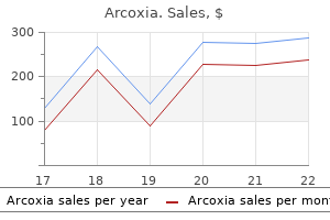
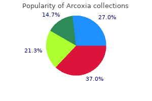
Buy generic arcoxia 90 mg
Posteriorly arthritis pain only on one side of body order arcoxia 120 mg on-line, where the vestibule ends arthritis reiki treatment arcoxia 90 mg generic with visa, the stratified squamous epithelium becomes thinner and undergoes a transition to the pseudostratified epithelium that characterizes the respiratory area. It is lined by the respiratory mucosa that incorporates a ciliated, pseudostratified columnar epithelium on its surface. The underlying lamina propria is firmly hooked up to the periosteum and perichondrium of the adjoining bone or cartilage. The medial wall of the respiratory area, the nasal septum, is smooth, however the lateral walls are thrown into folds by the presence of three shelf-like, bony projections referred to as conchae or turbinates. The conchae divide each nasal cavity into separate air chambers and play a twin function. They enhance floor space and trigger turbulence in airflow to permit extra efficient conditioning of inspired air. The ciliated, pseudostratified columnar epithelium of the respiratory mucosa is composed of 5 cell types: the epithelium of the respiratory area of the nasal cavity is essentially the same because the epithelium lining most of the parts that comply with within the conducting system. Because the respiratory epithelium of the trachea is studied and examined in desire to that of the nasal cavity, the above cell varieties are mentioned in the part on the trachea (page 670). The association of the vessels allows the inhaled air to be warmed by blood flowing via the part of the loop closest to the floor. The capillaries that reside close to the surface are arranged in rows; the blood flows perpendicular to the airflow, much as one would discover in a mechanical heatexchange system. These same vessels might become engorged and leaky throughout allergic reactions or viral infections such as the widespread chilly. The lamina propria then becomes distended with fluid, resulting in marked swelling of the mucous membrane with consequent restriction of the air passage, making breathing troublesome. Their secretions complement that of the goblet cells within the respiratory epithelium. By rising floor area, the conchae (turbinates) increase the efficiency with which the inspired air is warmed. The turbinates additionally enhance the efficiency of filtration of inspired air through the method of turbulent precipitation. Particulate matter suspended in the air stream is thrown out of the stream and adheres to the mucus-covered wall of the nasal cavity. Particles trapped on this layer of mucus are transported to the pharynx by the use of coordinated sweeping movements of cilia and are then swallowed. The lamina propria of the olfactory mucosa is instantly contiguous with the periosteum of the underlying bone (Plate 69, page 688). This connective tissue incorporates quite a few blood and lymphatic vessels, unmyelinated olfactory nerves, myelinated nerves, and olfactory glands. The olfactory epithelium, like the epithelium of the respiratory area, can also be pseudostratified, but it contains very totally different cell varieties. In residing tissue, this mucosa is distinguished by its slight yellowish brown color attributable to pigment in the olfactory epithelium and the associated olfactory glands. In humans, the entire floor space of the olfactory mucosa is simply about 10 cm2; in animals with an acute sense of odor, the whole floor area of the olfactory mucosa is � � � � Olfactory receptor cells are bipolar olfactory neurons that span the thickness of the epithelium and enter the central nervous system. Supporting or sustentacular cells are columnar cells which are similar to neuroglia cells and provide mechanical and metabolic support to the olfactory receptor cells. Basal cells are stem cells from which new olfactory receptor cells and supporting cells differentiate. This diagram shows the three main cell varieties positioned within the olfactory epithelium: the olfactory cell, supporting (sustentacular) cell, and basal cell. The olfactory cell is the receptor cell; it has an apical growth, the olfactory vesicle, from which lengthy, nonmotile cilia prolong. At its basal floor, it extends an axon into the connective tissue that joins with axons of other olfactory cells to form an olfactory nerve. The supporting cells, in distinction, are columnar and lengthen the complete thickness of the epithelium; their nuclei are positioned within the upper portion of the cell. Note that the ducts of the olfactory glands lengthen from the secretory portion of the gland to the epithelial surface. Supporting cells provide mechanical and metabolic support for the olfactory receptor cells. The cilia are normally as much as 200 m long and will overlap with cilia of adjoining olfactory receptor cells. The cilia are thought to be nonmotile, although some research means that they might have restricted motility. The basal area of the cell gives rise to an unmyelinated axonal course of that leaves the epithelial compartment. The collections of axons from olfactory receptor cells form the olfactory nerve (cranial nerve I). The olfactory axons are very fragile and may be harmed during traumatic head injury. They may be completely severed, resulting in anosmia (loss of the sense of smell). Autoradiographic research show that olfactory receptor cells have a life span of about 1 month. Olfactory receptor cells (and some neurons of the enteric division of the autonomic nervous system) seem to be the only neurons in the nervous system which may be readily replaced throughout postnatal life. Entire olfactory transduction pathways occur throughout the cilia of the olfactory receptor cells. Supporting cells are probably the most numerous cells within the olfac- Respiratory System tory epithelium. Adhering junctions are current between these cells and the olfactory receptor cells, however hole and tight junctions are absent. All the molecules that are concerned in olfactory transduction are positioned within long cilia that arise from the olfactory bulb. Olfactory receptors are specific for the olfactory receptor cells and belong to the household of G protein�coupled receptors (known as Golf). Thus, the olfactory system must decode olfactory impulses not from only the olfactory epithelium additionally contains cells present in much smaller numbers, known as brush cells. As famous, these cells are present within the epithelium of other components of the conducting air passages. The basal floor of a brush cell is in synaptic contact with nerve fibers that penetrate the basal lamina. The nerve fibers are terminal branches of the trigeminal nerve (cranial nerve V) that perform in general sensation rather than olfaction. Brush cells appear to be involved in transduction of basic sensory stimulation of the mucosa. In addition, presence of a microvillous border, vesicles near the apical cell membrane, and a well-defined Golgi equipment suggest that brush cells might be concerned in an absorptive in addition to a secretory function. Their nuclei are incessantly invaginated and lie at a level beneath those of the olfactory receptor cell nuclei.
Trusted arcoxia 60 mg
Glycogen particles appear at the periphery of the lipid droplets arthritis pain relief weight loss discount 60 mg arcoxia otc, and pinocytotic vesicles and basal lamina turn out to be more obvious arthritis blisters generic arcoxia 120 mg mastercard. The mature adipocyte is characterised by a single, giant lipid inclusion surrounded by a skinny rim of cytoplasm. White adipocytes derive from undifferentiated perivascular mesenchymal stem cells related to the adventitia of small venules. Brown adipocytes also have a mesenchymal origin; nonetheless, they derive from frequent skeletal myogenic progenitor cells present in dermatomyotomes of developing embryos. Lipoblasts develop an exterior (basal) lamina and start to accumulate quite a few lipid droplets in their cytoplasm. In white adipose tissue, these droplets fuse to type a single large lipid droplet that in the end fills the mature cell, compressing the nucleus, cytoplasm, and cytoplasmic organelles into a thin rim around the droplet. In the late stage of differentiation, the cells improve in size and turn into more spherical. Small lipid droplets coalesce to type a single giant lipid droplet that occupies the central portion of the cytoplasm. Eventually, the lipid mass compresses the nucleus to an eccentric position, producing a signet-ring look in hematoxylin and eosin (H&E) preparations. Structure of Adipocytes and Adipose Tissue Unilocular adipocytes are massive cells, generally 100 m or extra in diameter. Lipoblasts initially develop from stromal-vascular cells along the small blood vessels in the fetus and are freed from lipids. The nucleus is flattened and displaced to one facet of the lipid mass; the cytoplasm forms a thin rim around the lipid. The thin strand of meshwork that separates adjoining adipocytes represents the cytoplasm of each cells and the extracellular matrix. Photomicrograph of white adipose tissue, showing its attribute meshwork in an H&E� stained paraffin preparation. Each space represents a single giant drop of lipid earlier than its dissolution from the cell during tissue preparation. The surrounding eosin-stained materials represents the cytoplasm of the adjoining cells and a few intervening connective tissue. Highpower photomicrograph of a glutaraldehyde-preserved, plastic-embedded specimen of white adipose tissue. The cytoplasm of the person adipose cells is recognizable in some areas, and part of the nucleus of one of many cells is included within the aircraft of section. Because of the large size of adipose cells, the nucleus is occasionally noticed in a given cell. Adipose tissue is richly supplied with blood vessels, and capillaries are found on the angles of the meshwork where adjacent adipocytes meet. Special stains also reveal the presence of unmyelinated nerve fibers and quite a few mast cells. Regulation of Adipose Tissue It is nearly impossible to separate regulation of adipose tissue from digestive processes and features of the central nervous system. This layer separates the hydrophobic contents of the lipid droplet from the hydrophilic cytoplasmic matrix. The amount of adipose tissue in an individual is regulated by two physiologic systems. The first system, which is associated with short-term weight regulation, controls urge for food and metabolism on a day by day basis. The second system, which is associated with long-term weight regulation, controls urge for food and metabolism on a continuing basis (over months and years). The cytoplasm of the adipose cells reveals mitochondria (M) and glycogen (the latter appears as the very darkish particles). Each cell is separated by a slim space containing exterior (basal) lamina and an especially attenuated means of a fibroblast. Two hormones, leptin and insulin, are liable for longterm regulation of physique weight. The recently found potent urge for food stimulant ghrelin is a small, 28-amino-acid polypeptide produced by gastric epithelial cells. In addition to its appetite stimulatory role, it acts on the anterior lobe of the pituitary gland to launch development hormone. In humans, ghrelin features through receptors located in the hypothalamus, growing the sense of hunger. A genetic mutation in chromosome 15 causes Prader-Willi syndrome, during which an overproduction of ghrelin leads to morbid weight problems. In individuals with this syndrome, compulsive eating and an obsession with meals normally arise at an early age. The urge to eat in these individuals is physiologic, overwhelming, and very difficult to management. If not treated, these people usually die before age 30 of issues attributable to obesity. In experimental animal fashions, the addition of recombinant leptin to overweight, leptin-deficient ob/ob mice causes them to reduce their meals consumption and lose about 30% of their total physique weight after 2 weeks of remedy. Recent scientific findings point out that leptin most likely protects the body in opposition to weight loss in occasions of food deprivation. Antiobesity drug research is currently focusing on substances that may inhibit insulin and leptin signaling in the hypothalamus. Deposition and mobilization of lipid are influenced by neural and hormonal components. When adipose tissue is stimulated by neural or hormonal mechanisms, triglycerides are broken down into glycerol and fatty acids, a process referred to as mobilization. Neural mobilization is particularly necessary in periods of fasting and exposure to severe cold. During the early levels of experimental starvation in rodents, adipose cells in a denervated fats pad proceed to deposit fats. It is now recognized that norepinephrine (which is liberated by the endings of nerve cells of the sympathetic nervous system) initiates a collection of metabolic steps that lead to the activation of lipase. This enzyme splits triglycerides, which represent greater than 90% of the lipids stored in the adipocyte. Hormonal mobilization involves a complex system of hormones and enzymes that controls fatty-acid release from adipocytes. Insulin is a vital hormone that promotes lipid synthesis by stimulating lipid synthesis enzymes (fatty-acid synthase, acetyl-CoA carboxylase) and suppresses lipid degradation by inhibiting the action of hormone-sensitive lipase and thus blocking the discharge of fatty acids. Glucagon, another pancreatic hormone, and development hormone from the pituitary gland both improve lipid utilization (lipolysis). Brown adipose tissue, a key thermogenic tissue, is Insulin, the pancreatic hormone that regulates blood glucose levels, can be involved in regulation of adipose tissue metabolism. It enhances the conversion of glucose into the triglycerides of the lipid droplet by the adipocyte. Like leptin, insulin regulates weight by performing on mind centers in the hypothalamus.
Diseases
- Pierre Robin sequence faciodigital anomaly
- Limb reduction defect
- Immunodeficiency, secondary
- Shellfish poisoning, paralytic (PSP)
- Polyomavirus Infections
- Charcot Marie Tooth disease, neuronal, type B
- D-Glyceric acidemia
- Chromosome 10, monosomy 10q
- Bronchitis, Chronic
Arcoxia 60 mg proven
Frequently arthritis turmeric buy generic arcoxia 120 mg on line, when cell boundaries are noticed at still higher magnification (inset) arthritis pain back order arcoxia 90 mg line, a really small circular or oval profile is noticed halfway along the boundary. The cells that line the sinusoids (S) show little, if any, cytoplasmic detail in routine preparations. The endothelial cell, in distinction, is a squamous cell that has a smaller, attenuated, or elongated nucleus. Note that the wall of the vein is strengthened by connective tissue, largely collagen, which seems as homogeneous eosin-stained materials (asterisks). Hepatic sinusoids, liver, rat, glutaraldehyde�osmium fixation, toluidine blue 900. This figure shows a plastic-embedded liver specimen fastened by the method normally used for electron microscopy. In contrast to the H&E�stained preparation, it demonstrates to advantage the cytologic element of the hepatocytes and the sinusoids (S). This is glycogen that has been retained by the glutaraldehyde fixation and stained metachromatically by toluidine blue. Also evident are lipid droplets (L) of various measurement which have been preserved and stained black by the osmium used because the secondary fixative. The quantities of lipid and glycogen are variable and, beneath regular situations, replicate dietary intake. Examination of the hepatocyte cytoplasm also reveals small, punctate, dark-blue our bodies contrasted in opposition to the lighter blue background of the cell. They appear as empty round profiles when cross-sectioned and as elongate channels (lower right) when longitudinally sectioned. The floor of the Kupffer cell displays a very irregular or jagged contour because of the quite a few processes that present the cell with an extensive floor area. The bile is concentrated by the lively transport of salt from the bile and the passive movement of water in response to the salt transport. The mucosa is characterised by a tall columnar absorptive epithelium that intently resembles that of the gut and the colon in both its morphology and function. The epithelial cells are characterized by quite a few brief apical microvilli, apical junctional complexes, concentrations of mitochondria within the apical and basal cytoplasm, and complicated lateral plications. It consists of a mucosa (Muc), muscularis (Mus), and adventitia (Adv) and, on its free floor (not shown), a serosa. The mucosa is thrown into quite a few folds which might be notably pronounced when the muscularis is highly contracted. This is the same old histologic look of the gallbladder except, of course, steps are taken to fix and preserve it in a distended state. Occasionally, the part cuts via a recess in a fold, and the recess could then resemble a gland (arrows). The epithelium has characteristics that distinguish it from the absorptive epithelium of different organs, such as the intestines. Only one cell kind, tall columnar cells, is present within the epithelial layer (see upper proper figure). This is related to its absorptive perform and is in distinction to the staining of cells which are engaged within the production of protein. Lastly, with respect to its absorptive operate, the epithelial cells incessantly exhibit distended intercellular areas at their basal side (see upper right figure arrows). This is a function related to the transport of fluid across the epithelium and, as noted above, generally seen in intestinal absorptive cells. These are readily obvious in figure on top left and a part of the wall of the Rokitansky-Aschoff sinus is proven at larger magnification in determine below. This specimen was taken from a website near the neck of the gallbladder the place mucous glands are sometimes present. Note the attribute flattened nuclei at the base of the cell and the frivolously stained appearance of the cytoplasm, options attribute of mucin-secreting cells. It is a combined gland containing both an exocrine component and an endocrine part which have distinctive characteristics. The exocrine element is a compound tubuloacinar gland with a branching network of ducts that convey the exocrine secretions to the duodenum. The endocrine part is isolated as extremely vascularized islets of epithelioid tissue (islets of Langerhans). The islet cells secrete a wide range of polypeptide and protein hormones, most notably insulin and glucagon, which regulate glucose metabolism all through the opposite tissues of the physique. Other hormones secreted by islet cells embody somatostatin, pancreatic polypeptide, vasoactive intestinal peptide, secretin, motilin, and substance P. All of these substances, aside from insulin, are additionally secreted by the inhabitants of enteroendocrine cells within the gut, the organ from which the pancreas is derived throughout embryonic development. While insulin and glucagon act primarily in endocrine regulation of distant cells, the opposite hormones (and glucagon) have vital roles within the paracrine regulation of the insulin-secreting B cells of the pancreatic islet. The pancreas is surrounded by a fragile capsule of reasonably dense connective tissue. Also throughout the lobule are the small blood vessels and the connective tissue serving as a stroma for the parenchymal elements of the gland. The lumen of the acinus is small, and only in fortuitous sections by way of an acinus is the lumen included (asterisks). Some acini reveal a centrally positioned cell with cytoplasm that exhibits no particular staining characteristics in H&E�stained paraffin sections. This determine demonstrates particularly well the morphology and relationships of the intercalated ducts. Note, first, the cross-sectioned intralobular duct (InD) consisting of cuboidal epithelium. This is because the aircraft of part cuts mainly through the cells somewhat than the lumen. As a consequence, this figure offers a great view of the nuclei of the duct cells. In addition, they show a staining pattern just like that of centroacinar cells and totally different from that of nuclei of the parenchymal cells. Three principal features are carried out by this method: air conduction, air filtration, and gasoline exchange (respiration). In addition, air passing by way of the larynx is used to produce speech, and air passing over the olfactory mucosa within the nasal cavities carries stimuli for the sense of odor. The respiratory system additionally participates to a lesser degree in endocrine capabilities (hormone manufacturing and secretion), in addition to regulation of immune responses to inhaled antigens. Lungs develop from the laryngotracheal diverticulum of the foregut endoderm and its surrounding thoracic splanchnic mesenchyme. This preliminary diverticulum grows into the thoracic splanchnic mesenchyme surrounding the foregut. This lung bud divides into the left and right bronchial buds, which enlarge to kind the primordium of the left and proper primary bronchi. Bronchial buds together with the encircling thoracic mesenchyme differentiate into lobar bronchi with subsequent progressive divisions into segmental bronchi. Each segmental bronchus with its surrounding mesenchyme further differentiates and divides to kind the bronchopulmonary segments of the lung.
Buy arcoxia 120 mg amex
An instance of this modification happens inside the microvillus psoriatic arthritis in my feet arcoxia 90 mg buy line, where actin filaments are cross-linked by the actin-bundling proteins fascin and fimbrin arthritis in feet toes symptoms arcoxia 60 mg generic on line. Actin-capping proteins block additional addition of actin molecules by binding to the free end of an actin filament. An instance is tropomodulin, which may be isolated from skeletal and cardiac muscle cells. Tropomodulin binds to the free end of actin myofilaments, regulating the size of the filaments in a sarcomere. Actin cross-linking proteins are responsible for cross-linking actin filaments with each other. Immunofluorescence micrograph of a chick cardiac myocyte stained for actin (green) to present the thin filaments and for tropomodulin (red) to present the location of the slow-growing ends of the thin filaments. Tropomodulin seems as regular striations due to the uniform lengths and alignment of the thin filaments in sarcomeres. The polarity of the skinny filament is indicated by the fast-growing end and the slow-growing finish. The troponin advanced binds to every tropomyosin molecule each seven actin monomers alongside the length of the skinny filament. Extensive studies have revealed the presence of quite so much of other nonmuscle myosin isoforms which are answerable for motor features in lots of specialised cells, such as melanocytes, kidney and intestinal absorptive cells, nerve development cones, and inner ear hair cells. As in lamellipodia, these protrusions comprise free aggregations of 10 to 20 actin filaments organized in the same course, again with their plus ends directed towards the plasma membrane. In listeriosis, an infection caused by Listeria monocytogenes, the actin polymerization machinery of the cell can be hijacked by the invading pathogen and utilized for its intracellular movement and dissemination throughout the tissue. Actin polymerization permits bacteria to move right into a neighboring cell by forming protrusions within the host plasma membrane. Intermediate Filaments Intermediate filaments play a supporting or general structural function. These rope-like filaments are referred to as intermediate as a result of their diameter of 8 to 10 nm is between those of actin filaments and microtubules. Nearly all intermediate filaments encompass subunits with a molecular weight of about 50 kDa. Some proof means that lots of the steady structural proteins in intermediate filaments advanced from highly conserved enzymes, with solely minor genetic modification. Intermediate filaments are shaped from nonpolar and extremely variable intermediate filament subunits. Locomotion is achieved by the force exerted by actin filaments by polymerization at their growing ends. This mechanism is used in many migrating cells-in specific, on reworked cells of invasive tumors. As a results of actin polymerization at their forefront, cells extend processes from their surface by pushing the plasma membrane ahead of the growing actin filaments. The modern extensions of a crawling cell are called lamellipodia; they contain elongating organized bundles of actin filaments with their plus ends directed towards the plasma membrane. These processes can be observed in plenty of other cells that exhibit small protrusions Unlike those of microfilaments and microtubules, the protein subunits of intermediate filaments present considerable variety and tissue specificity. The long, straight actin filament cores or rootlets (R) extending from the microvilli are cross-linked by a dense network of actin filaments containing numerous actin-binding proteins. The community of intermediate filaments may be seen beneath the terminal web anchoring the actin filaments of the microvilli. Mechanism of brush border contractility studied by the quick-freeze, deep-etch method. Intermediate filaments are assembled from a pair of helical monomers that twist round each other to form coiled-coil dimers. Each tetramer, appearing as a person unit, is aligned along the axis of the filament. The ends of the tetramers are certain collectively to type the free ends of the filament. This meeting process provides a steady, staggered, helical array in which filaments are packed together and additionally stabilized by lateral binding interactions between adjoining tetramers. Intermediate filaments are a heterogeneous group of cytoskeletal parts present in numerous cell varieties. Intermediate filaments are self-assembled from a pair of monomers that twist around one another in parallel fashion to form a steady dimer. Two coiled-coil dimers then twist round one another in antiparallel fashion to generate a staggered tetramer of two coiled-coil dimers. Each tetramer, performing as a person unit, aligns along the axis of the filament and binds to the free end of the elongating structure. This staggered helical array is moreover stabilized by lateral binding interactions between adjoining tetramers. These are probably the most numerous teams of intermediate filaments and are referred to as keratins (cytokeratins). Keratins solely assemble as heteropolymers; an acid cytokeratin (class 1) and a primary cytokeratin (class 2) molecule type a heterodimer. Each keratin pair is attribute of a specific type of epithelium; however, some epithelial cells may specific multiple pair. According to new nomenclature, keratins are divided into three expression teams: keratins of simple epithelia, keratins of stratified epithelia, and structural keratins, � � � � additionally referred to as exhausting keratins. Keratin filaments span the cytoplasm of epithelial cells and, through desmosomes, join with keratin filaments in neighboring cells. They characterize a various family of cytoplasmic filaments discovered in many cell varieties. In contrast to keratins, class 3 proteins (with the exception of desmin) preferentially form homopolymeric filaments containing only one type of intermediate protein. Historically, this group has been called neurofilaments; they contain intermediate filament proteins which are expressed largely in axons of nerve cells. All three proteins form neurofilaments that extend from the cell body into the ends of axons and dendrites, providing structural help. However, genes for class four proteins additionally encode several different intermediate filament proteins. These embody nestin and -internexin in nerve cells in addition to synemin, syncoilin, and paranemin in muscle cells. In distinction to other forms of intermediate filaments found within the cytoplasm, lamins are positioned inside the nucleoplasm of just about all differentiated cells within the body. This is a lens-specific group of intermediate filament, or "beaded filaments" containing two proteins, phakinin and filensin. The periodic bead-like surface appearance of those filaments is attributed to the globular structure of the carboxy-terminus of the filensin molecule, which projects out from the assembled filament core. Distribution of vimentin (red) and actin filaments (green) is shown in cultured fibroblasts from human fetal lung. Vimentin is an intermediate filament protein expressed in all cells of mesenchymal origin. In cultured fibroblasts, vimentin filaments are seen centrally within the cell cytoplasm, whereas the actin filaments are aggregated primary close to the cell floor.

Generic arcoxia 120 mg line
Several different varieties of T lymphocytes have been recognized: cytotoxic arthritis diet natural news buy 90 mg arcoxia free shipping, helper arthritis exercises buy discount arcoxia 60 mg online, suppressor, and gamma/delta. The suppressor T cells may also perform in suppressing B-cell differentiation and in regulating erythroid cell maturation within the bone marrow. Their location inside the pores and skin and mucosa of inner organs permits them to operate in the first line of defense towards invading organisms. Monocytes Monocytes are the precursors of the cells of the mononuclear phagocytotic system. Immunolabeling methods have made it possible to establish particular forms of T cells and examine their perform. Regulatory (suppressor) T cells represent a phenotypically numerous population of T lymphocytes that may functionally suppress an immune response to international and self-antigen by influencing the exercise of different cells in the immune system. They travel from the bone marrow to the body tissues, where they differentiate into the assorted phagocytes of the mononuclear phagocytotic system-that is, connective tissue macrophages, osteoclasts, alveolar macrophages, perisinusoidal macrophages in the liver (Kupffer cells), and macrophages of lymph nodes, spleen, and bone marrow amongst others (see Chapter 6, Connective Tissue). The indentation is the location of the cell middle the place the well-developed Golgi apparatus and centrioles are located. Monocytes additionally include clean endoplasmic reticulum, rough endoplasmic reticulum, and small mitochondria. Although these cells are categorised as agranular, they include small, dense, azurophilic granules. These granules include typical lysosomal enzymes just like these discovered in the azurophilic granules of neutrophils. Monocytes transform into macrophages, which function as antigen-presenting cells in the immune system. During irritation, the monocyte leaves the blood vessel at the site of inflammation, transforms right into a tissue macrophage, and phagocytoses micro organism, different cells, and tissue particles. The nucleus is markedly indented, and adjoining to this website, a centriole (C) and a quantity of other Golgi profiles (G) are evident. This electron micrograph exhibits a portion of a megakaryocyte from a bone marrow part. The cytoplasm reveals evidence of platelet formation as indicated by the intensive platelet demarcation channels. The "foamy" peripheral cytoplasm of the megakaryocyte represents areas during which segmentation to form platelets is happening. The membrane that lines these channels arises by invagination of the plasma membrane; subsequently, the channels are in continuity with the extracellular house. The continued growth and fusion of the platelet demarcation membranes outcome within the full partitioning of cytoplasmic fragments to type particular person platelets. After entry into the vascular system from the bone marrow, the platelets flow into as discoid structures about 2 to 3 m in diameter. Structurally, platelets may be divided into 4 zones based mostly on organization and function. The glycocalyx consists of glycoproteins, glycosaminoglycans, and a variety of other coagulation components adsorbed from the plasma. The structural zone, near the periphery, comprises microtubules, actin filaments, myosin, and actin-binding proteins that type a community supporting the plasma membrane. The marginal band containing eight to 24 coiled microtubules resides as a bundle immediately below the actin filament network. It consists of mitochondria, peroxisomes, glycogen particles, and at least three forms of granules dispersed inside the cytoplasm. The most quite a few granules are granules (300 to 500 nm in diameter) that include mainly fibrinogen, coagulation elements, plasminogen, plasminogen activator inhibitor, and platelet-derived growth issue. The contents of those granules play an necessary function within the preliminary section of vessel restore, blood coagulation, and platelet aggregation. High-magnification electron micrograph of a platelet located between an erythrocyte on the left and an endothelial cell on the right. Visible organelles embrace a mitochondrion, microtubules, a single profile of the surfaceconnected open canalicular system, profiles of the dense tubular system, the moderately dense granules, a single very dense granule, and glycogen particles. They facilitate platelet adhesion and vasoconstriction in the space of the injured vessel. The granules are similar to lysosomes present in other cells and comprise a number of hydrolytic enzymes. The contents of granules function in clot resorption through the later stages of vessel repair. In effect, open canaliculi are invaginations into the cytoplasm from the plasma membrane. They continuously survey the endothelial lining of blood vessels for gaps and breaks. When a blood vessel wall is injured or broken, the uncovered connective tissue on the damaged web site promotes platelet adhesion. Serotonin is a potent vasoconstrictor that causes the vascular easy muscle cells to contract, thereby reducing local blood move on the site of damage. The glycocalyx of the platelets offers a reaction floor for the conversion of soluble fibrinogen into fibrin. The initial platelet plug is remodeled right into a definitive clot generally known as a secondary hemostatic plug by further tissue factors secreted by the broken blood vessel. After the definitive clot is formed, platelets cause clot retraction, probably as a perform of the actin and myosin found within the structural zone of the platelet. Contraction of the clot permits the return of normal blood circulate through the vessel. High-magnification scanning electron micrograph reveals preliminary stage of blood clot formation. Red blood cells are entrapped in a free mesh of fibrin fibers that are extensively cross-linked to kind an impermeable hemostatic plug that prevents motion of cells and fluids from the lumen of the injured vessel. An additional role of platelets is to assist repair the injured tissues past the vessel itself. Platelet-derived growth factor released from the granules stimulates easy muscle cells and fibroblasts to divide and permit tissue repair. It provides relative numbers and calculations obtained from the cells (erythrocytes and leukocytes) and shaped elements (thrombocytes) within the blood sample. These calculations are normally performed by automated blood cell counters that analyze different elements of blood utilizing the precept of flow cytometry design. As a thin stream of fluid with suspended cells flows through slim tubing within the cell counter, the light detector and electrical impedance sensor establish totally different cell varieties based on their dimension and electrical resistance. Data obtained from automated blood analyzers were normally very correct because of the large variety of cells counted (10,000) in every category. However, in some circumstances, guide cell count underneath a light microscope continues to be necessary. Leukocyte count can be elevated after strenuous train due to stress, or in pregnancy and labor. Hyperleukocytosis (leukocyte count 100 109 cells/L) is usually a sign of leukemia (type of blood cancer). The main types of white blood cells reported are neutrophils, eosinophils, basophils, lymphocytes, and monocytes.
Discount arcoxia 120 mg otc
Some of the IgE binds to the plasma membranes of mast cells in the lamina propria (see pages 179�182) duramax for arthritis in dogs arcoxia 90 mg visa, selectively sensitizing them to specific antigens derived from the lumen arthritis in back lumbar cheap 60 mg arcoxia free shipping. Dimeric dIgA consists of two monomeric IgA subunits and a polypeptide J chain additionally produced by the plasma cell. In the lamina propria, dIgA binds to the polymeric immunoglobulin receptor (pIgR) on the basal cell membrane of the enterocyte. The pIgR�IgA complex enters the cell by endocytosis and is carried out throughout the endocytotic vesicles to the early endosomal compartment after which to the apical surface (a course of referred to as transcytosis). Endocytic vesicles fuse with the apical plasma membrane, the pIgR is proteolytically cleaved, and dIgA is released with the extracellular portion of the pIgR receptor. J chain Submucosa A distinguishing attribute of the duodenum is the presence of submucosal glands. The submucosa consists of a dense connective tissue and localized websites that include aggregates of adipose cells. This extremely alkaline secretion in all probability serves to protect the proximal small intestine by neutralizing the acidcontaining chyme delivered to it. It additionally brings the intestinal contents close to the optimal pH for the pancreatic enzymes that are additionally delivered to the duodenum. The dashed line marks the boundary between the villi and the standard intestinal glands (crypts of Lieberk�hn). These are branched tubular glands whose secretory element consists of columnar cells. This intestinal stem cell niche (zone of cell replication) is restricted to the lower one-half of the gland and contains extremely proliferative intermediate cells (as previously explained) and cells at varied stages of differentiation. A cell destined to become a goblet cell or absorptive cell often undergoes several additional divisions after it leaves the pool of stem cells. The epithelial cells migrate upward within the intestinal gland onto the villus the place they endure apoptosis and slough off into the lumen. Autoradiographic studies have shown that the renewal time for absorptive and goblet cells within the human small gut is four to 6 days. Enteroendocrine cells and Paneth cells are also derived from the stem cells at the base of the intestinal gland. They live for about 4 weeks and are then replaced by differentiation of a nearby "dedicated" cell in the intestinal gland. As talked about in the chapter on epithelial tissue (page 146), expression of the transcription factor Math1 seems to determine the fate of differentiating cells within the intestinal stem cell area of interest. Inhibition of Math1 expression characterizes the default developmental pathway into absorptive intestinal cells (enterocytes). The colon is additional subdivided on the premise of its anatomic location into ascending colon, transverse colon, descending colon, and sigmoid colon. Local contractions displace intestinal contents each proximally and distally; this sort of contraction is identified as segmentation. They serve to circulate the chyme locally, mixing it with digestive juices and shifting it into contact with the mucosa for absorption. Peristalsis, the second sort of contraction, involves coordinated motion of both round and longitudinal muscle layers and strikes the intestinal contents distally. Haustra coli which would possibly be seen sacculations between the teniae coli on the exterior floor of the cecum and colon. Omental appendices that are small fatty projections of the serosa, noticed on the outer floor of the colon. Serosa the serosa of the elements of the small gut that are situated intraperitoneally within the stomach cavity corresponds to the final description at the beginning of the chapter. Mucosa the mucosa of the large intestine has a "clean" floor; neither plicae circulares nor villi are present. The glands consist of simple columnar epithelium, as does the intestinal floor from which they invaginate. The morphology of absorptive cells is essentially identical to that of the enterocytes of the small gut. Elimination of semisolid to stable waste materials is facilitated by the large quantities of mucus secreted by the numerous goblet cells of the intestinal glands. The mucosal epithelium of the massive gut incorporates the identical cell varieties as the small gut besides Paneth cells, that are normally absent in people. This photograph exhibits the outer (serosal) surface (left) and inner (mucosal) floor (right) of the transverse colon. The smooth mucosal surface shows semilunar folds (arrows) fashioned in response to contractions of the muscularis externa. The ratio decreases, nevertheless, approaching 1:1, near the rectum, where the number of goblet cells increases. This photomicrograph of an H&E preparation exhibits the mucosa and part of the submucosa. The floor epithelium is steady with the straight, unbranched, tubular intestinal glands (crypts of Lieberk�hn). As the absorptive cells are adopted into the glands, they become fewer in quantity, whereas the goblet cells increase in number. The extremely cellular lamina propria incorporates quite a few lymphocytes and different cells of the immune system. Comparative electron-microscopic options of normal, hyperplastic, and adenomatous human colonic epithelium. They secrete mucus continuously, even to the purpose where they attain the luminal floor. Here, on the surface, the secretion price exceeds the synthesis rate, and "exhausted" goblet cells appear within the epithelium. These cells are tall and thin and have a small number of mucinogen granules in the central apical cytoplasm. An occasionally observed cell type, the caveolated "tuft" cell, has also been described within the colonic epithelium; however, this cell could additionally be a form of exhausted goblet cell. The turnover times of the epithelial cells of the massive intestine are much like these of the small gut. Senile epithelial cells that attain the mucosal surface bear apoptosis and are shed into the lumen at the midpoint between two adjacent intestinal glands. Lamina Propria Although the lamina propria of the large intestine incorporates the identical basic elements as the the rest of the digestive tract, it demonstrates some extra structural options and higher improvement of some others. These embrace the following: � Collagen table, which represents a thick layer of col- Epithelial Cell Renewal in the Large Intestine All intestinal epithelial cells in the large gut derive from a single stem cell inhabitants. As within the small intestine, all the mucosal epithelial cells of the large gut arise from stem cells situated at the backside of the intestinal gland. The intermediate cell sorts discovered in the � lagen and proteoglycans that lies between the basal lamina of the epithelium and that of the fenestrated absorptive venous capillaries.
Shiitake (Shiitake Mushroom). Arcoxia.
- How does Shiitake Mushroom work?
- Dosing considerations for Shiitake Mushroom.
- Prostate cancer.
- What is Shiitake Mushroom?
- Reducing high cholesterol and other conditions.
Source: http://www.rxlist.com/script/main/art.asp?articlekey=96669
Arcoxia 120 mg mastercard
The high endothelial venule is recognized by its endothelium arthritis medication starting with d buy generic arcoxia 60 mg, which is composed of cells that are cuboidal arthritis pain barometric pressure buy cheap arcoxia 90 mg. A cross-sectioned profile of a postcapillary venule is shown in the inset at greater magnification (700). The endothelial cell nuclei are spherical and are flippantly stained, in contrast to the nuclei of the encircling lymphocytes, which are of comparable measurement and form however are densely stained. This vessel additionally reveals three lymphocytes (arrows) that are within the means of migrating via the wall of the vessel. These cells wrap across the collagen bundles that type the supporting trabecular framework of the node. In H&E preparations, these characteristics allow for the excellence between the reticular cell and the lymphocyte. The openings in the vessel wall (arrows) are sites in which the medullary sinuses are emptying their contents into the lymphatic vessel. The substance of the spleen, the splenic pulp, consists of purple pulp and white pulp, so named because of their look in contemporary tissue. The red pulp incorporates giant numbers of red blood cells that it filters and degrades. Aged, broken, or irregular red blood cells are trapped by macrophages associated with uncommon vascular sinuses within the pink pulp. These macrophages break down the purple cells, begin the metabolic breakdown of hemoglobin, and retrieve and retailer the iron from the heme for reutilization within the formation of latest purple blood cells within the bone marrow. The white pulp, however, is so named as a result of its content material of lymphocytes seems in life as whitish areas. In tissue sections, nonetheless, the nuclei of the intently packed lymphocytes impart an overall blue-staining response. The lymphatic tissue that constitutes the white pulp differs from nodules seen elsewhere in that it follows and ensheathes a blood vessel, the central artery. The lymphatic tissue surrounding the artery reveals periodic expansion, thus forming the nodules. This figure reveals, at the next magnification, the pink pulp and a portion of the trabecular vein from the realm enclosed in the uppermost rectangle within the high determine. In this specimen, the venous sinuses could be seen to benefit because the red blood cells within the sinuses have lysed and appear as unstained "ghosts"; solely the nuclei of the white cells are readily seen. The wall of the vein is thin, however the trabecula (T) containing the vessel provides the appearance of being part of the vessel wall. In humans in addition to in different mammals, the capsule and the trabeculae that stretch from the capsule comprise myofibroblasts. Under situations of increasing physical stress, contraction of those cells will occur and trigger fast expulsion of blood from the venous sinuses into the trabecular veins and, thus, into the final circulation. This determine reveals, at greater magnification, the splenic nodule in the rectangle in the proper portion of the figure above. Small arterial vessels and capillaries, branches of the central artery, provide the white pulp, and a few move into the reticular community of the marginal zone, terminating in a funnel-shaped orifice. Venous sinuses are additionally discovered within the marginal zone, and infrequently, arterial vessels may open into the sinuses. The particulars of the vascular provide are, at greatest, difficult to resolve in typical H&E preparations. The penicillar arterioles, the terminal branches of the central artery, provide the purple pulp but are likewise difficult to resolve. Thus, the comparatively clear areas with scattered nuclei characterize the lumen of the venous sinus; the nuclei are these of white blood cells. In this specimen, the pink blood cells have been lysed leaving only a clear define Red pulp, spleen, human, H&E 1,200. This micrograph is a high magnification of the area within the rectangle of the earlier micrograph. Other than the lysed red blood cells, which appear as empty round profiles, numerous lymphocytes (Ly) are current in the lumen. A slim but clearly visible intercellular space is current between adjacent cells. Also, processes of macrophages positioned outside of the sinuses within the splenic cords prolong between the endothelial cells and into the lumen of the sinuses to monitor the passing blood for international antigens. A macrophage (M), recognized by residual our bodies in its cytoplasm, is seen just outdoors of the sinus. At the highest of the micrograph, two venous sinuses (arrows) could be seen emptying into the trabecular vein. These small trabecular veins converge into larger veins, which ultimately unite giving rise to the splenic vein. The structural parts that are stained by the silver within the nodule consist of reticular fibers. The fantastic, thread-like stained material that encircles the venous sinuses is a ordinary modification of basement membrane. Where the vessel has been minimize deeper along its long axis, the basement membrane appears as dot-like structures (arrowheads). A threedimensional reconstruction of the basement membrane would reveal it as a collection of ring-like buildings. The supporting reticular stroma arises from endodermal epithelium and produces a mobile reticulum. Lymphocytes come to lie within the interstices of the cellular reticulum, and these two mobile elements, the lymphocytes and the epithelioreticular cells, comprise the bulk of the organ. The stem lymphocytes that migrate into the endodermal rudiment within the embryo derive from the yolk sac and, later, from the pink bone marrow. These lymphocytes proliferate and turn into immunologically competent within the thymus, differentiating into the thymus-dependent lymphocytes. Some of those lymphocytes migrate to different tissues to populate the thymus-dependent parts of lymph nodes and spleen as properly as to reside within the unfastened connective tissue. Many lymphocytes die or are destroyed within the thymus because in the random process by which they purchase the flexibility to acknowledge and react to antigens, they become programmed towards "self" antigens. A blood�thymus barrier is shaped by the sheathing of the perivascular connective tissue of the thymus by the epithelioreticular cells. The thymus involutes during adolescence and is often difficult to recognize within the grownup. A connective tissue capsule (Cap) surrounds every lobe of the 2 lobes of the thymus and sends trabeculae (T) into the parenchyma to kind lobules. Examination of the thymus at low magnification reveals the lobules (L) composed of a dark-staining basophilic cortex (C) and a lighter staining and comparatively eosinophilic medulla (M). The cortex accommodates quite a few densely packed lymphocytes, whereas the medulla incorporates fewer lymphocytes and is consequently much less densely packed. It is the relative difference within the lymphocyte inhabitants (per unit area) and, in particular, the staining of their nuclei with hematoxylin that creates the difference in look between cortex (C) and medulla (M). Note that some of the medullary areas bear a resemblance to germinal facilities of different lymphatic organs because of the medulla appearing as isolated circular areas (upper left of prime figure). The medullary component, nevertheless, is actually a continuous branching mass surrounded by cortical tissue.
60 mg arcoxia order with amex
The subendothelial M layer of the intima consists of connective tissue with both collagen and elastic fibers rheumatoid arthritis icd 9 discount 90 mg arcoxia amex. The external border of the intima is bounded by an inner elastic membrane A that represents the first layer of the many concentric fenestrated laminae within the I media of the vessel arthritis urica definition 120 mg arcoxia discount visa. At roughly 50 years, individual laminae begin to present indicators of degeneration and gradually turn out to be replaced by M collagen resulting in a gradual loss of elasticity of the aortic wall. The adventitia consists of irregular dense connective tissue with interA mixed elastic fibers that are inclined to be organized in a circumferential sample. The adventitia (A) incorporates an abundance of collagenous fibers and stains more densely than that of both the media or intima. The lower micrograph is from an adult and has been stained to reveal the elastic part of the vessel wall. The intima (I) could be very lightly stained, on this case, because of the paucity of elastic material. The media (M) is closely stained as a outcome of the presence of huge amounts of elastic laminae. The adventitia (A) incorporates in addition to the dense connective tissue a average amount of elastic fibers. The inset shows the intima at higher magnification and consists of part of the media. The specimen proven here has been stained to distinguish collagen from elastic materials. The media (M) contains quite a few elastic lamellae that appear because the black wavy traces. Careful examination of the media reveals nuclei of smooth muscle cells dispersed between the elastic lamellae. This micrograph shows the outer portion of the media (M) with its elastic lamellae. The outer portion of the adventitia incorporates numerous elastic fibers which appear as the black, dot-like structures. These elastic fibers are arranged in a circumferential sample, thus when sectioned, they seem as black, dot-like constructions. Thus, because the arterial tree is traced further from the guts, the elastic tissue is considerably lowered and easy muscle turns into the predominant element of the tunica media. The muscular arteries are characterised, nonetheless, by a refractile inner elastic membrane separating the tunica intima from the tunica media and, usually, by an external elastic membrane separating the tunica media from the tunica adventitia. Muscular arteries, or arteries of medium caliber, constitute nearly all of the named arteries in the body. The veins have the identical three layers of their walls, but the tunica media is thinner than in the accompanying artery, and the tunica adventitia is the predominant layer within the wall. In this photomicrograph, the lumen of the artery is on the left and the lumen of the vein is on the right. The collagen bundles of the unfastened connective tissue beneath the tunica adventitia are bigger than these of the adventitia, and there are fewer cells on this portion of the specimen. Arterioles have an endothelial lining and clean muscle within the wall, however the easy muscle is limited in thickness to one or two cells. There may or will not be an internal elastic membrane, according to the scale of the vessel. In the traditional relationship between an arteriole and a capillary community, contraction of the smooth muscle of the arteriole wall reduces or shuts off the blood going to the capillaries. A precapillary sphincter is formed by a slight thickening of the sleek muscle at the origin of a capillary mattress from an arteriole. The arteriole on the left is recognized as a big arteriole, based on the presence of two discrete layers of smooth muscle cells that form the tunica media of the vessel. The nuclei of the muscle cells appear in longitudinal profile as a result of the circumferential arrangement of the cells. The endothelial cell nuclei of the vessel appear as small spherical profiles surrounding the lumen. These cells are elongate and oriented with their long axis in the course of move. The arteriole on the best is a very small arteriole, having solely a single layer of easy muscle. The endothelial cell nuclei appear because the small round profiles at the luminal surface. A venule is seen in proximity to the larger arteriole, and a cross-section of peripheral nerve (N) is seen in proximity to the smaller arteriole. Compare the wall of the venule, consisting only of endothelium and a thin layer of connective tissue, with the arterioles. After the arteriole makes an acute flip (segment numbered 2), the vessel wall is cut to reveal the lumen. Here, the graceful muscle nuclei seem as round profiles and the nuclei of the endothelial cells lining the lumen appear in longitudinal profile. In the section numbered 4, the cut is deeper, again displaying the lumen and some of the endothelial cells in face view (arrowheads). Because of its twisting path by way of the part, its wall has been cut such that the one layer of muscle cells of the tunica media is seen in different planes along its length. In the segment numbered 1, on the left, the vessel wall has been cut tangentially. The lymphatic vessel shown in this figure exhibits a area where the vessel is making a U-shaped turn in the plane of the part, thus disappearing on the high and backside of the micrograph. The wall of the vessel consists of an endothelial lining and a small quantity of connective tissue, with one being indistinguishable from the opposite. A valve (Val), which is attribute of lymphatic vessels, is seen within the vessel. The arrows indicate nuclei which would possibly be just barely visible at this magnification; most of them belong to endothelial cells. Typically, the lumen incorporates precipitated lymph materials (L); sometimes, lymphocytes could also be present. A venule (V) can be present; it could readily be distinguished from the lymphatic vessel by the presence of red blood cells in the lumen. The lymphatic system consists of groups of cells, tissues, and organs that monitor body surfaces and internal fluid compartments and react to the presence of potentially harmful substances. Lymphocytes are the definitive cell kind of the lymphatic system and the effector cells within the response of the immune system to dangerous substances. The various lymphatic organs and lymphatic tissues are sometimes collectively referred to as the immune system. Lymphatic tissues serve as websites the place lymphocytes proliferate, differentiate, and mature. These at the moment are immunocompetent cells that can distinguish between "self " (molecules usually present within an organism) and "nonself " (foreign molecules-i. The body is continually uncovered to pathogenic (disease-causing) organisms and dangerous substances from the external environment (infectious microorganisms, toxins, and international cells and tissues). In addition, adjustments might occur in cells (such as transformation of normal cells into cancerous cells) that give them characteristics of international cells.

Arcoxia 90 mg low cost
Regenerating motor axons are stained green for neurofilaments; reestablished connections with two neuromuscular junctions are visualized in pink colour arthritis in the feet exercises arcoxia 60 mg discount with visa, which displays specific staining for postsynaptic acetylcholine receptors; Schwann cells are stained blue for S100 arthritis in hands arcoxia 90 mg cheap with amex, which represents a Schwann cell�specific calcium-binding protein. Regenerating axons extended alongside Schwann cells, which led them to the unique synaptic sites of the muscle fibers. They migrate to the positioning of nerve harm, proliferate, after which phagocytize myelin particles. When an axon is injured, the blood�nerve barrier (see page 389) is disrupted alongside the complete length of the injured axon, which permits for the inflow of these cells into the location of damage. The presence of huge numbers of monocytederived macrophages speeds up the process of myelin elimination, which in peripheral nerves is usually accomplished within 2 weeks. Another issue that affects nerve regeneration is the formation of a glial (astrocyte-derived) scar that fills the empty area left by degenerated axons. These processes contain not solely neurons but in addition supportive cells such as Schwann cells and oligodendrocytes in addition to phagocytic cells corresponding to macrophages and microglia. This permits large infiltration of monocyte-derived macrophages, which are liable for the process of myelin removing. Rapid clearance of myelin debris allows for axon regeneration and subsequent restoration of the blood�nerve barrier. They divide and bear marked hypertrophy with a visible increase in the number of their cytoplasmic processes. This course of is referred to as reactive gliosis, while the ensuing permanent scar is most often referred to as a plaque. Several organic mechanisms for induction and maintenance of reactive gliosis have been proposed. These reactive microglial cells migrate toward the positioning of damage and exhibit marked phagocytic exercise. However, their phagocytic exercise and ability to remove myelin particles is much lower than that of monocyte-derived macrophages. The coverage of traumatic degeneration depends on the severity of the injury and often extends for only one or a quantity of internodal segments. Sometimes, traumatic degeneration extends more proximally than one or a number of nodes of Ranvier and should result in demise of the cell body. Retrograde signaling to the cell physique of an injured nerve causes a change in gene expression that initiates reorganization of the perinuclear cytoplasm. Axonal injury also initiates retrograde signaling to the nerve cell physique resulting in the upregulation of a gene referred to as c-jun. C-jun transcription issue is concerned in early in addition to later stages of nerve regeneration. Reorganization of the perinuclear cytoplasm and organelles begins inside a few days. Initially, Nissl our bodies disappear from the center of the neuron and transfer to the periphery of the neuron in a course of referred to as chromatolysis. The modifications within the cell physique are proportional to the quantity of axoplasm destroyed by the injury; intensive loss of axoplasm can result in demise of the cell. Before the event of modern dyes and radioisotope tracer strategies, Wallerian degeneration and chromatolysis have been used as research tools. These tools allowed researchers to hint the pathways and destination of axons and the localization of the cell bodies of experimentally injured nerves. Cellular bands information the growth of new nerve processes (neurites or sprouts) of regenerating axons. A progress cone develops in the distal portion of every sprout that consists of filopodia rich in actin filaments. The suggestions of the filopodia set up a direction for the development of the growth cone. They preferentially interact with proteins of the extracellular matrix corresponding to fibronectin and laminin discovered within the exterior lamina of the Schwann cell. Thus, if a sprout associates itself with a band of Bungner, it regenerates between the layers of exterior lamina of the Schwann cell. After crossing the site of damage, sprouts enter the surviving cellular bands in the distal stump. Axonal regeneration results in Schwann cell redifferentiation, which occurs in a proximal-to-distal course. Redifferentiated Schwann cells upregulate genes for myelin-specific proteins and downregulate c-jun. If physical contact is reestablished between a motor neuron and its muscle, function is normally reestablished. As mentioned above, division of dedifferentiated Schwann cells is the first step in the regeneration of a severed or crushed peripheral nerve. Initially, these cells arrange themselves in a sequence of cylinders referred to as endoneurial tubes. Removal of myelin and axonal debris from inside the tubes causes them to eventually collapse. Proliferating Schwann cells organize Microsurgical techniques that rapidly reestablish intimate apposition of severed nerve and vessel ends have made reattachment of severed limbs and digits, with subsequent reestablishment of operate, a relatively frequent process. Clinically, traumatic neuroma normally appears as a freely movable nodule on the website of nerve harm and is characterised by ache, particularly on palpation. Traumatic neuroma of the injured motor nerve prevents reinnervation of the affected muscle. In myelinated nerves, Schwann cells produce the myelin sheath from compacted layers of their very own cell membranes which may be wrapped concentrically across the nerve cell course of. The junction between two adjacent Schwann cells known as the node of Ranvier and is where electrical impulse is regenerated for high-speed propagation along the axon. In unmyelinated nerves, nerve processes are enveloped in the cytoplasm of Schwann cells. Each neuron consists of a cell physique or perikaryon (contains the nucleus, Nissl bodies, and different organelles), an axon (usually the longest means of the cell physique; transmits impulses away from the cell body), and a variety of other dendrites (shorter processes that transmit impulses towards the cell body). Neurons communicate with other neurons and with effector cells by specialised junctions known as synapses. The most common sort of synapses is chemical synapses, in which neurotransmitters are released from a presynaptic neuron and bind to receptors situated on the postsynaptic neuron (or goal cell). A chemical synapse has a presynaptic factor (filled with synaptic vesicles containing neurotransmitter), a synaptic cleft (separates the presynaptic neuron from the postsynaptic neuron), and a postsynaptic membrane (containing receptors for neurotransmitter). It is protected by with specialized nerve endings (synapses) and ganglia containing nerve cell our bodies. Individual nerve fibers are held together by connective tissue organized into endoneurium (surrounds each individual nerve fiber and associated Schwann cell), perineurium (surrounds each nerve fascicle), and epineurium (surrounds a peripheral nerve and fills the spaces between nerve fascicles).
Purchase arcoxia 60 mg on-line
It is usually identified by the presence of aggregates of rounded chondrocytes (isogenous groups) among bundles of collagen fibers and by the basophilic staining of the capsular matrix material and territorial matrix secreted by these cells arthritis relief for dogs aspirin arcoxia 60 mg cheap otc. The tissue has a fibrous appearance arthritis in birds feet arcoxia 60 mg discount with amex, and at this low magnification, the nuclei of the fibroblasts (F) appear as small, elongate, or spindle-shaped bodies. There are comparatively few fibroblasts present, as is attribute of dense connective tissue. Some of the chondrocytes appear as elongate clusters of cells, whereas others appear in single-file rows. The matrix materials immediately surrounding the chondrocytes has a homogeneous look and is, thereby, distinguishable from the fibrous connective tissue. Cartilage matrix material could be detected greatest by observing the bigger group of chondrocytes at the left of this determine and then observing this identical area within the figure above. This figure shows the world circumscribed by the rectangle in the micrograph above at greater magnification. The chondrocytes are contained inside lacunae (arrows), and their cytoplasm stains deeply. Bone is a specialised form of connective tissue that, like different connective tissues, consists of cells and extracellular matrix. The function that distinguishes bone from different connective tissues is the mineralization of its matrix, which produces an especially onerous tissue able to providing support and safety. By advantage of its mineral content, bone also serves as a storage website for calcium and phosphate. Both calcium and phosphate can be mobilized from the bone matrix and taken up by the blood as wanted to preserve acceptable levels all through the body. Thus, along with help and safety, bone plays an necessary secondary function in the homeostatic regulation of blood calcium levels. Bone matrix contains mainly sort I collagen together with other matrix (noncollagenous) proteins. All collagen molecules constitute about 90% of the whole weight of the bone matrix proteins. The matrix also incorporates different matrix (noncollagenous) proteins that constitute the ground substance of bone. Both the collagen and the bottom substance turn into mineralized to type bone tissue. The four main teams of noncollagenous proteins discovered within the bone matrix are the next: � Proteoglycan macromolecules include a core protein the main structural component of bone matrix is type I collagen and, to a lesser extent, type V collagen. Trace � with various numbers of covalently hooked up aspect chains of glycosaminoglycans (hyaluronan, chondroitin sulfate, and keratan sulfate). They are also liable for binding development components and may inhibit mineralization. Multiadhesive glycoproteins are liable for attachment of bone cells and collagen fibers to the mineralized ground substance. Typically, a bone consists of bone tissue and other connective tissues, including hemopoietic tissue, fats tissue, blood vessels, and nerves. If the bone types a freely movable joint, otherwise referred to as a synovial joint, hyaline cartilage is current. The capability of the bone to perform its skeletal operate is attributable to the bone tissue, ligaments and, where current, the articular (hyaline) cartilage. Canaliculi course by way of the mineralized matrix, connecting adjoining lacunae and allowing contact between the cell processes of neighboring osteocytes (Plate 11, page 244). In this manner, a steady community of canaliculi and lacunae-containing cells and their processes is formed all through the whole mass of mineralized tissue. Osteoclasts are bone-resorbing cells current on bone surfaces where bone is being eliminated or transformed (reorganized) or where bone has been damaged. This photograph shows a longitudinally sectioned proximal epiphysis of the femur after the bone was processed by alkaline hydrolysis. The inside of the bone reveals a spongy configuration and represents spongy (cancellous) bone. It consists of quite a few interconnecting bony trabeculae separated by a labyrinth of interconnecting marrow areas. The outer portion of the bone has a strong structure and represents compact (dense) bone. It is particularly well seen in the diaphysis, which encloses bone marrow cavity. Inset from the rectangular area reveals enlargement of interphase between spongy and compact bone. Osteoprogenitor cells and osteoblasts are developmental precursors of the osteocyte. The spaces throughout the meshwork are continuous and, in a residing bone, are occupied by marrow and blood vessels. Bones are classified in accordance with form; the location of spongy and compact bone varies with bone form. It is beneficial, then, to define briefly the kinds of bones and survey the place the two kinds of bone tissue are located. On the basis of form, bones can be categorized into four teams: calvaria [skull cap] and the sternum). They consist of two layers of comparatively thick compact bone with an intervening layer of spongy bone. The flared portion of the bone between the diaphysis and the epiphysis is known as the metaphysis. A large cavity filled with bone marrow, called the marrow or medullary cavity, varieties the internal portion of the bone. In the shaft, nearly the complete thickness of the bone tissue is compact; at most, only a small quantity of spongy bone faces the marrow cavity. Short bones possess a shell of compact bone and have spongy bone and a marrow area on the inside. Short bones often kind movable joints with their neighbors; like lengthy bones, their articular surfaces are lined with hyaline cartilage. Elsewhere, periosteum, a fibrous connective tissue capsule, covers the outer surface of the bone. Bone Outer Surface of Bones Bones are covered by periosteum, a sheath of dense fibrous connective tissue containing osteoprogenitor cells. The diaphysis (shaft) of an extended bone in the grownup contains yellow bone marrow in a large marrow cavity surrounded by a thick-walled tube of compact bone. The proximal and distal ends, or epiphyses, of the lengthy bone consist mainly of spongy bone with a thin outer shell of compact bone. The expanded or flared part of the diaphysis nearest the epiphysis is referred to because the metaphysis. Except for the articular surfaces which would possibly be covered by hyaline (articular) cartilage, indicated in blue, the outer surface of the bone is roofed by a fibrous layer of connective tissue known as the periosteum. Bones are lined by a periosteum besides in areas where they articulate with another bone. The periosteum that covers an actively growing bone consists of an outer fibrous layer that resembles other dense connective tissues and an inside, more cellular layer that contains the osteoprogenitor cells.
